REQUEST AN APPOINTMENT OR BOOK A CONSULANT – Sargam.dange.18@gmail.com
This article addresses hip dislocation that results from a traumatic injury. To learn about pediatric developmental hip dislocation, please read Developmental Dislocation (Dysplasia) of the Hip (DDH). To learn about dislocation after total hip replacement, please read Total Hip Replacement.
A traumatic hip dislocation occurs when the head of the thighbone (femur) is forced out of its socket in the hip bone (pelvis). It typically takes a major force to dislocate the hip. Car collisions and falls from significant heights are common causes and, as a result, other injuries like broken bones often occur with the dislocation.
A hip dislocation is a serious medical emergency. Immediate treatment is necessary.
Description
When there is a hip dislocation, the femoral head is pushed either backward out of the socket, or forward.
Posterior dislocation. In approximately 90% of hip dislocation patients, the thighbone is pushed out of the socket in a backwards direction. This is called a posterior dislocation. A posterior dislocation leaves the lower leg in a fixed position, with the knee and foot rotated in toward the middle of the body.
Anterior dislocation. When the thighbone slips out of its socket in a forward direction, the hip will be bent only slightly, and the leg will rotate out and away from the middle of the body.
When the hip dislocates, the ligaments, labrum, muscles, and other soft tissues holding the bones in place are often damaged, as well. The nerves around the hip may also be injured.
Symptoms
A hip dislocation is very painful. Patients are unable to move the leg and, if there is nerve damage, may not have any feeling in the foot or ankle area.
Cause
Motor vehicle collisions are the most common cause of traumatic hip dislocations. The dislocation often occurs when the knee hits the dashboard in a collision. This force drives the thigh backwards, which drives the ball head of the femur out of the hip socket. Wearing a seatbelt can greatly reduce your risk of hip dislocation during a collision.
A fall from a significant height (such as from a ladder) or an industrial accident can also generate enough force to dislocate a hip.
With hip dislocations, there are often other related injuries, such as fractures in the pelvis and legs, and back, abdominal, knee, and head injuries. Perhaps the most common fracture occurs when the head of the femur hits and breaks off the back part of the hip socket during the injury. This is called a posterior wall acetabular fracture-dislocation.
REQUEST AN APPOINTMENT OR BOOK A CONSULANT – Sargam.dange.18@gmail.com
Diagnosis
To diagnose a dislocated hip or other source of hip pain, an orthopedist will conduct a physical exam and order imaging of the hip in the form of an X-ray, MRI and/or CT scan.
Doctor Examination
A hip dislocation is a medical emergency. Call for help immediately. Do not try to move the injured person, but keep him or her warm with blankets.
In cases in which hip dislocation is the only injury, an orthopaedic surgeon can often diagnose it simply by looking at the position of the leg. Because hip dislocations often occur with additional injuries, your doctor will complete a thorough physical evaluation.
Your doctor may order imaging tests, such as x-rays, to show the exact position of the dislocated bones, as well as any additional fractures in the hip or thighbone.
Complications
A hip dislocation can have long-term consequences, particularly if there are associated fractures.
Nerve injury. As the thighbone is pushed out of the socket, particularly in posterior dislocations, it can crush and stretch nerves in the hip. The sciatic nerve, which extends from the lower back down the back of the legs, is the nerve most commonly affected. Injury to the sciatic nerve may cause weakness in the lower leg and affect the ability to move the knee, ankle and foot normally. Sciatic nerve injury occurs in approximately 10% of hip dislocation patients. The majority of these patients will experience some nerve recovery.
Osteonecrosis. As the thighbone is pushed out of the socket, it can tear blood vessels and nerves. When blood supply to the bone is lost, the bone can die, resulting in osteonecrosis (also called avascular necrosis). This is a painful condition that can ultimately lead to the destruction of the hip joint and arthritis.
Arthritis. The protective cartilage covering the bone may also be damaged, which increases the risk of developing arthritis in the joint. Arthritis can eventually lead to the need for other procedures, like a total hip replacement.
Recovery
It takes time—sometimes 2 to 3 months—for the hip to heal after a dislocation. The rehabilitation time may be longer if there are additional fractures. The doctor may recommend limiting hip motion for several weeks to protect the hip from dislocating again. Physical therapy is often recommended during recovery.
Patients often begin walking with crutches within a short time. Walking aids, such as walkers, crutches and, eventually, canes, help patients get mobilized.
Treatment
Reduction Procedures
If there are no other injuries, the doctor will administer an anesthetic or a sedative and manipulate the bones back into their proper position. This is called a reduction.
In some cases, the reduction must be done in the operating room with anesthesia. In rare cases, torn soft tissues or small bony fragments block the bone from going back into the socket. When this occurs, surgery is required to remove the loose tissues and correctly position the bones.
Following reduction, the surgeon will request another set of x-rays and possibly a computed tomography (CT) scan to make sure that the bones are in the proper position.
Hip dislocations after a total hip replacement
Hip dislocations in people who have had a total hip replacement (THR) are relatively infrequent among otherwise healthy people who follow the precautions provided by their orthopedic surgeon and physical therapist. But higher rates of dislocations occur in certain hip replacement patients: the elderly, those with other physical disabilities, those who had a THR after a hip fracture or after other hip surgeries, and in those who had one or more hip dislocations prior to a THR (for example, if muscles and ligaments around the hip were disrupted from the prior dislocation and weakened as a result). If a patient experiences multiple dislocations after THR, he or she is usually a good candidate for a hip revision surgery.
REQUEST AN APPOINTMENT OR BOOK A CONSULANT – Sargam.dange.18@gmail.com
REQUEST AN APPOINTMENT OR BOOK A CONSULANT – Sargam.dange.18@gmail.com
Hand fractures are common in the general population with relative propensity seen in contact-sport athletes (For example, boxers, football players) and manual laborers,
A metacarpal fracture
Is a break in one of the five metacarpal bones of either hand.
Are categorized as being fractures of the head, neck, shaft, and base (from distal at the metacarpal phalangeal joint to proximal
at the wrist).
Boxer fracture is another name for a fracture of the fourth or fifth metacarpal, one of the most common metacarpal fractures.
The mechanisms of these injuries vary from axial loading forces to direct blows to the dorsal hand
Clinically Relevant Anatomy
The metacarpals are long, thin bones that are located between the carpal bones in the wrist and the phalanges in the digits.
Each is comprised of a base, shaft, and head.
The proximal bases of the metacarpals articulate with the carpal bones,
Distal heads of the metacarpals articulate with the proximal phalanges and form the knuckles.
The 1st metacarpal is the thickest and shortest of these bones.
The 3rd metacarpal is distinguished by a styloid process on the lateral side of its base.
Soft tissues generally involved with fractures include cartilage, joint capsule, ligaments, fascia, and the dorsal hood fibers.
With severe polytrauma cases, the tendons and nerves adjacent to the fracture can also be injured.
Etiology
Metacarpal fractures typically occur secondary to a direct blow or fall directly onto the hand.
These fractures commonly occur during athletic activities, particularly in contact sports. Almost one-fourth of cases occur during athletic events.
Sporting injury is frequently the cause among younger patients
Work-related injuries are often the cause in middle-aged patients
Falls are typically the cause of the elderly.
Fifth metacarpal fractures often occur secondary to punching a wall or other solid object (hence the eponym, “boxer’s fracture”)
Hand Fractures
Makeup about 40% of all acute hand injuries
Constitute about 20% of all fractures occurring below the elbow
Metacarpal Fractures
Typically occur in patients aged 10-40 years
Men are more likely to be affected than women.
Young men sustain metacarpal fractures secondary to a punching mechanism or a direct blow to the hand
Geriatric females sustain these injuries secondary to a low energy fall.
The incidence rate of fracture seen in association with each digit’s metacarpal bone increases from the radial to the ulnar side.
The incidence rate of 2nd metacarpal fractures is lower than the incidence rate of 5th metacarpal fractures.
Bennett fracture is the most common fracture involving the base of the thumb. This fracture refers to an intraarticular fracture that separates the palmar ulnar aspect of the first metacarpal base from the remaining first metacarpal.
The fractures of the metacarpals can be divided into three parts.
The first, neck fractures, occurs often when a person punches another person or object. In the majority of cases, surgical intervention is not essential to treat this condition.
The metacarpal shaft fractures are often produced by longitudinal compression, torsion, or direct impact. They are described by the appearance of their respective fracture patterns and can be divided by transverse, oblique, spiral, and comminuted.
Metacarpal base fractures are rare and have a minimal consequence because the motion of the joint is small. More common are the fractures of the base of the fifth digit and are the result of a longitudinally directed force
REQUEST AN APPOINTMENT OR BOOK A CONSULANT – Sargam.dange.18@gmail.com
Characteristics/Clinical Presentation
Patients with metacarpal fractures generally present with
Pain
Swelling
Ecchymosis (bruise)
Limitation of movement
Deformity. Knuckle asymmetry may be observed, and the knuckle may appear to be missing.
Finger misalignment may also be noted.
A metacarpal head fracture is associated with axial compression of the extended digit which causes severe discomfort.
In a metacarpal base fracture, movement of the wrist or longitudinal compression exacerbates the pain.
Any metacarpal fracture angulation can produce a pseudo-claw deformity.
Differential Diagnosis
Injuries to neighboring bones (carpal bones, phalanges) and associated soft tissues (ligaments, tendons) need to be excluded.
Evaluation
The evaluation includes:
Standard radiographs of the hand (anteroposterior, lateral, and oblique). In the vast majority of cases, this will be enough to confirm the diagnosis and form a management plan. Confirmation of more subtle injuries can be obtained using special views such as Brewerton (metacarpal heads), Roberts, and Betts (thumb) views.
CT is sometimes necessary for the base of metacarpal fractures to check for any intra-articular displacement and determine if there is a need for surgery
Outcome Measures
Grip Strength: measured with a dynamometer
Range of motion
Patient Specific Functional Scale
DASH
Michigan Hand Outcome Questionnaire (MHO): In this questionnaire, they assess 6 criteria for people with a hand disorder: overall hand function, activities of daily living (ADL), pain, work performance, aesthetics, and patient satisfaction with hand function.
Medical Management
The goal of treatment is a restoration of anatomy and function.
Antibiotics and tetanus prophylaxis are options for open fractures as per standardized guidelines.
The modality of treatment will vary depending on skin integrity (open versus closed fracture), the number of digits/metacarpals fractured, the stability of the specific, degree of comminution, displacement, and/or rotational malalignment
In general, increasing degrees of displacement, comminution, and rotational malalignment are critical factors in assessing the fracture patterns potential for stability and reduction maintenance with nonoperative management.
The GP/Specialist after assessing the fracture will perform gentle tests and imaging to work out if surgery is needed.
If surgery is not needed a physiotherapist will make a custom splint, which will support the healing fracture.
Physical Therapy Management
Full strength and range of motion is the goal of rehabilitation.
Under the physiotherapist’s instructions
Hand exercises with light resistance such as rubber bands or squeeze ball can help if there is scarring or extensor lag develops.
Soft tissue recovery may be more of a problem than the bony one.
Rest and elevation are important, and so is the quality of splinting – poor splinting can cause stiffness, pressure sores, or even compartment syndrome
REQUEST AN APPOINTMENT OR BOOK A CONSULANT – Sargam.dange.18@gmail.com
Physiotherapists use a number of techniques to regain movement in the hand, wrist, and fingers, including:
Swelling management with massage and compression garments
Soft tissue massage to help with muscle tension and pain
Providing clients with a home exercise program of specific movements and strengthening exercises.
Most hand fractures can be treated non-operatively
More specific Advice.
These are the steps to be followed in a stable fracture:
one or other of the below stabilizing techniques could be used
Buddy strapping the injured digit to another digit is used as a non-operative technique. This is used with or without the application of varying degrees of splint. The ‘buddy’ reduces the risk of rotational deformity.
The splinting of the fracture should be: 20 degrees wrist extension; MCP joint 60-70 degree flexion and IP joint extension
Early motion is generally considered appropriate when there are stable fractures or rigid fractures.
Generally, AROM (active ROM) exercises without resistance can begin 2 to 3 weeks after operative treatment in uninvolved or bordering/adjacent joints.
Active Motion: If the fracture is internally fixed, the active range of motion can start early. Most fractures are treated by immobilization, but the active motion can begin after three weeks of therapy, starting with the joints not splintered during the initial immobilization. This phase usually lasts 3-6 weeks. Specific tendon gliding should be included in the active motion.
Tendon gliding is important to prevent adhesions, increased circulation about the fracture site, decreased edema and compression at the fracture site.
Exercises For Tendon Gliding
Claw posture to achieve extensor digitorum communis tendon glide over the metacarpal bone
Intrinsic plus posture to achieve central slip. Lateral bands glide over proximal phalanx 1
Flexor digitorum profundus (FDP) blocking exercises to glide FDP tendon over the phalanx
Hook fist posture to promote selective FDP tendon glide
Flexor digitorum sublimis blocking exercise to glide FDS tendon over middle phalanx
Sublimis fist posture to promote selective FDS tendon glide
Passive Motion
Passive motion can be initiated after sufficient clinical healing at approximately 5-6 weeks of therapy.
The timing of initiation of joint mobilization depends on the structures involved in the injury. If the structures resisting the force are not involved in the injury, joint mobilization can be initiated at the same time as active motion. Compression on the fracture can result in shortening, angulation or rotational malalignment of the bone.
Traditional PROM aims to assist in articular cartilage healing, reduce swelling, and stiffness.
Resistive Motion: Four weeks after the injury light resistance can be performed in most metacarpal fractures which are treated by immobilization. Active motion should only be continued if healing has not started.
Resistive exercise should also be delayed when a fracture is fixed by pinning until these pins are removed, to ensure the stability of the fracture. Light resistive exercise helps with scar remodeling and improved motion. There are several types of resistive exercises such as the weight-well exercises. This kind of exercise strengthens the finger flexors (FDP and FDS muscles).
Functional activities and work simulation should be included in the resistive exercises as soon as possible.
Conclusion
Main points on metacarpal fractures:
Common hand injury
Require thorough assessment consisting of the history, examination, and radiological investigations
They mostly divide into open or closed, based on the digit they affect, intra-articular or extra-articular status, and based on the location on the bone itself (head, neck, shaft, base)
May have conservative or operative treatment
Can have long-term sequelae requiring further management
Rehabilitation goals are return of full strength and range of motion.
Rest and elevation are important, and so is the quality of splinting – poor splinting can cause stiffness, pressure sores, or even compartment syndrome.
Physiotherapy is an critical element in the restoration of good hand function.
REQUEST AN APPOINTMENT OR BOOK A CONSULANT – Sargam.dange.18@gmail.com
The scaphoid bone is the most commonly fractured carpal bone. In young children and the elderly population, scaphoid fractures are rare. The scaphoid bone is stronger than the relatively weak distal radius in these age groups.
A delay in diagnosis of scaphoid fractures can lead to a variety of adverse outcomes that include nonunion (no consolidation), delayed union, decreased grip strength, and range of motion, along with osteoarthritis of the radiocarpal joint. Timely diagnosis, appropriate immobilization, and referral to surgical opinion when indicated can decrease the likelihood of adverse outcomes.
Scaphoid fractures are common, but present unique challenges because of the particular geometry of the fractures and the tenuous vascular pattern of the scaphoid. Delays in diagnosis and inadequate treatment for acute scaphoid fractures can lead to non-unions and subsequent degenerative wrist arthritis.
Epidemiology
Scaphoid fractures predominantly affect young adults, with a mean age of 29 years.
Higher incidence in males.
Unusual in the pediatrics population and the elderly population where the physis or distal radius, respectively, are more likely to fracture first.
Scaphoid fractures account for 15% of acute wrist injuries.
Scaphoid fractures have a high incidence of nonunion (8-10%), frequent malunion, and late sequelae of carpal instability and post-traumatic arthritis.
No blood vessels enter the proximal pole of the scaphoid, thus a higher incidence of aseptic necrosis and nonunion is noted with fractures on this side of the scaphoid
Displaced fractures frequently are associated with ligamentus tears in the wrist.
Characteristics/ Clinical Presentation
Patients typically present with wrist pain following a fall onto an outstretched hand.
Axial loading of the wrist with it in forced hyperextension and radial deviation can cause the fracture as the scaphoid impacts on the dorsal rim of the radius.
Traumatic injury through contact sports and road traffic accidents are also common causes.
Following the traumatic event there will be a massive force of the hand on the arm through the scaphoid bone
The symptoms will likely be:
Deep, dull ache in the radial part of the wrist.
Aggravated pain by pinching and gripping.
Localised wrist swelling with fullness in the anatomical snuffbox
Localised bruising
Tenderness on palpation of the radial side of the wrist
Differential Diagnosis
These diagnoses can be differentiated by the location of tenderness, pain with certain maneuvers, and radiographic abnormalities.
Distal radius fracture
Other carpal bone fractures
Scapholunate dissociation
De Quervain’s tenosynovitis
Osteoarthritis
Tendinopathy
Diagnostic Procedures
Plain X-rays are commonly used to diagnose the fracture, but this approach may miss up to 16% of fractures in the absence of clear-cut lucent lines on plain radiographs plain radiographs have 64% specificity for scaphoid fractures.
Research has shown that the use of MRI or CTs effective in indeterminate a scaphoid fracture. Both methods have been shown to detect fractures, but the MRI found some significant ligamentous and carpal instabilities in addition to the scaphoid fracture.
Outcome Measures
DASH or QuickDASH (Disabilities of Arm, Shoulder or Hand)
PRWE, a fifteen-item questionnaire was designed to measure wrist pain and disability.
PEM (Patient Evaluation Measure) has a simple layout with questions asked in a visual analog form. Patients are asked to read and comprehend the question alone and not the description of each interval answer.
Assessment
Subjective assessment
History of trauma i.e. fall onto an outstretched hand
Dull pain which is aggravated by hand and wrist movements
Restricted thumb range of movement
Objective exam
When examining a patient with a suspected scaphoid injury, it is important to compare the injured wrist with the uninjured wrist.
Presentation may include:
Anatomical snuffbox tenderness on examination – highly sensitive 90% indication of scaphoid fracture, but it is nonspecific 40%
Tenderness of the scaphoid tubercle: the physician extends the patient’s wrist with one hand and applies pressure to the tuberosity at the proximal wrist crease with the opposite hand. This provides better diagnostic information; sensitivity 87%, specificity 57%
Pain with the scaphoid compression test (i.e. axially/longitudinally compressing a patient’s thumb along the line of the first metacarpal) was shown to be helpful in identifying a scaphoid fracture, but in another study
Pain in the snuffbox with pronation of the wrist followed by ulnar deviation (52% percent positive predictive value, 100% percent negative predictive value)
Medical Management
Suspected fractures with positive clinical findings on examination but negative radiographs should have a follow-up with films repeated in 7-14 days. If pain persists and radiographs are still normal, then further imaging in the form of MRI or CT should be undertaken. Pain management with the assistance of pharmacists should be considered.
Surgical Management
Indications for operative management include:
Displacement greater than 1mm
An intrascaphoid angle greater than 35 degrees (humpback deformity)
A radiolunate angle of more than 15 degrees
Transcaphoid perilunate dislocation
Proximal pole fractures
Comminuted fractures
Non displaced waist fractures in individuals that need to return quickly to work/sport
Nonunion or avascular necrosis
Surgical fixation involves the insertion of a single or multiple screws and can be done percutaneously or via an open procedure. The latter is preferable for non-unions and those fractures that exhibit gross displacement with the former for acute, minimally displaced fractures.
Conservative management
Fractures that are non-displaced and within the distal third of the bone can be managed non-operatively with immobilization in a cast. Debate exists as to whether a long or short arm cast is optimal and whether a thumb spica should be included to immobilize the thumb, no evidence currently suggests one option is better than the other.
Six weeks immobilization is normally required with repeat radiographs taken at this time to assess for the union.
Time to union varies depending on the location of the fracture.
The distal-third would be expected to heal within 6-8 weeks for approximately 90% of non-displaced or minimally displaced (≤ 0.5 mm) scaphoid waist fractures,
middle-third within 8-12 weeks
proximal third within weeks.
Scaphoid waist fractures with moderate displacement (0.5-1.5 mm) can be treated conservatively, require prolonged cast immobilization for eight to ten weeks.
The relative increase in time to healing while moving from distal to proximal is secondary to the tenuous blood supply and retrograde arterial flow.
As a basic rule, in a patient with a clinically suspected scaphoid fracture but negative initial radiographs, it is reasonable to apply a short arm thumb spica and re-evaluate the patient in two weeks. If a cast is not applied, the fracture can worsen over the following months. At the two-week visit, the patient should be free of pain, and a follow-up radiograph should be obtained.
Types of fracture
The fractured scaphoid exhibits certain behavior that inhibits healing. Fracture fragments are inherently unstable and prone to displacement and require motionless contact to achieve union. As mentioned before, the blood supply of the scaphoid is tenuous.
For therapeutic decision making, the scaphoid is divided into three anatomic sections: proximal, medial, and distal (see image). Fractures are further subdivided into displaced and non-displaced types.
Non-displaced Fractures
Non-displaced distal fractures heal well with strict immobilization in a well-molded short arm thumb spica. Controversy exists over whether to use a long arm or a short arm cast. he current treatment for this type of fracture is a thumb spica, but some evidence suggests that the thumb could be omitted from the cast.
Screw fixation may speed recovery to pre-injury activities; referral for surgery may be indicated, depending on the needs of the patient.
As the fracture line moves proximally, there is more risk of displacement and nonunion; therefore, it would be appropriate to refer these patients for orthopedic consultation. If conservative treatment is attempted, a long arm cast with thumb immobilization is appropriate.
Displaced Fractures
Fractures with even small amounts of displacement are prone to nonunion, and operative treatment is recommended.
For the fixation, double-threaded headless screws are preferred. Which operative technique to use depends on the fracture morphology. Splinting and referral are indicated.
Traditionally, un-displaced and stable scaphoid fractures are treated by casting in short- or long-arm casts.
Physiotherapy management
After the period of immobilization either post-operatively or conservatively, once the fracture is considered stable and cast removed it is likely the hand and wrist will be stiff and have reduced muscle strength.
The primary goals of physiotherapy are:
Restore active range of movement (AROM)
Reduce swelling
Increase grip and wrist strength
Return to functional goals and tasks
ROM exercises
ROM exercises in the initial stages after immobilization should focus on active-assisted ROM as the hand and wrist will be stiff.
These exercises should focus on the wrist and thumb, however, the fingers, elbow and shoulder also need to be considered as after immobilization these may also be stiff.
If full ROM is still restricted it may be useful in the therapy session to do manual therapy in the form of joint mobilisations to the radio-carpal joint, radio-ulnar joint and potentially to the carpal joints.
Other forms of manual therapy may also be helpful to reduce any residual swelling or pain such as soft tissue work or massage techniques.
Strengthening exercises
Once a full or functional AROM has been restored it is essential to undergo strengthening exercises of the wrist and hand.
This is an essential step in rehabilitation as without strengthening the hand long standing functional deficits may be present and also put the patient at risk of further injury.
Functional restoration
After full AROM has been restored and a good baseline strength has been regained focus should turn to individualized goals and tasks.
The demographic of scaphoid fractures tend to be younger to middle-aged people therefore it is likely they will be active or have jobs or family to attend to. So specific rehab and exercises need to be individualized to meet these goals and expectations. Full function will eventually be restored if the fracture has been appropriately managed in the initial stages i.e. no missed avascular necrosis.
In the sporting population it has been shown that early surgical intervention led to quicker return to play approximately 6-11 weeks versus 4-16 weeks for conservative management.
REQUEST AN APPOINTMENT OR BOOK A CONSULANT – Sargam.dange.18@gmail.com
Ankle injuries are often thought of as sports injuries. But you don’t have to be an athlete or even a “weekend warrior” to turn your ankle and hurt it. Something as simple as walking on an uneven surface can cause a painful, debilitating sprain.
Ankle injuries can happen to anyone at any age. However, men between 15 and 24 years old have higher rates of ankle sprain, compared to women older than age 30 who have higher rates than men. Half of all ankle sprains occur during an athletic activity. Every day in the U.S., 25,000 people sprain their ankle. And more than 1 million people visit emergency rooms each year because of ankle injuries. The most common ankle injuries are sprains and fractures, which involve ligaments and bones in the ankle. But you can also tear or strain a tendon.
At one time or another, everyone has had a minor toe, foot, or ankle injury that caused pain or swelling. Most of the time our body movements do not cause problems, but it’s not surprising that symptoms develop from everyday wear and tear, overuse, or an injury.
Toe, foot, or ankle injuries most commonly occur during:
Sports or recreational activities.
Work-related tasks.
Work or projects around the home.
In children, most toe, foot, or ankle injuries occur during sports, play, or falls. The risk for injury is higher in sports with jumping, such as basketball, or sports with quick direction change, such as soccer or football. Any bone injury near a joint may injure the growth plate (physis) in a child and needs to be evaluated.
Certain athletes, such as dancers, gymnasts, or soccer or basketball players, have an increased risk of toe, foot, or ankle injuries.
Older adults are at higher risk for injuries and fractures because they lose muscle mass and bone strength as they age. They also have more problems with vision and balance, which increases their risk of injury.
Most minor injuries will heal on their own, and home treatment is usually all that is needed to relieve your symptoms and promote healing.
Causes
An ankle injury occurs when the ankle joint is twisted too far out of its normal position. Most ankle injuries occur either during sports activities or while walking on an uneven surface that forces the foot and ankle into an unnatural position. The unnatural position of the ankle in high-heeled shoes or walking in unstable, loose-fitting clogs or sandals is also a factor that may contribute to ankle injuries. In addition to wearing faulty footwear, an ankle injury can happen as a result of:
Tripping or falling
Landing awkwardly after a jump
Walking or running on uneven surfaces
A sudden impact such as a car crash
Twisting or rotating the ankle
Rolling the ankle
Different Signs for Different Ankle Injuries
The symptoms of a sprain and of a fracture are very similar. In fact, fractures can sometimes be mistaken for sprains. That’s why it’s important to have an ankle injury evaluated by a doctor as soon as possible. The signs include:
Pain, often sudden and severe
Swelling
Bruising
Inability to walk or bear weight on the injured joint
With a sprain, the ankle may also be stiff. With a fracture the area will be tender to the touch, and the ankle may also look deformed or out of place.
If the sprain is mild, the swelling and pain may be slight. But with a severe sprain, there is much swelling and the pain is typically intense.
Tendinitis and acute tears of the peroneal tendon result in both pain and swelling. In addition, the ankle area will feel warm to the touch with tendinitis. With an acute tear, there will be a weakness or instability of the foot and ankle.
Sporadic pain on the outside of the ankle
Weakness or instability in the ankle
An increase in the height of the foot’s arch
With the subluxation you will notice ankle instability or weakness. You also may notice sporadic pain behind the outside ankle bone and a “snapping” feeling around the ankle bone.
REQUEST AN APPOINTMENT OR BOOK A CONSULANT – Sargam.dange.18@gmail.com
Diagnosis
The first thing a doctor will do is ask questions about how the injury occurred. Then the doctor will examine the ankle, noting the amount of swelling and bruising. The physical examination of the ankle may be painful because the doctor needs to move the ankle to evaluate the pain and swelling in order to make a proper diagnosis.
The doctor may order an ankle X-ray to determine whether there are any broken bones. In addition to an ankle X-ray, your doctor may ask for X-rays of the leg and foot to determine whether there may be other related injuries. If the doctor suspects a stress fracture, the doctor will ask for other imaging scans such as an MRI, which will show more detail about the injury. If there is a fracture, the doctor may also ask for a stress test, which is a special X-ray taken with pressure applied to the joint. This will help the doctor determine whether surgery is needed.
For most ankle injuries, pain is controlled by using an over-the-counter medication such as acetaminophen or other nonsteroidal anti-inflammatory drug such as ibuprofen. The specific treatment of the injury depends on the type of injury.
What Should Someone Do After an Ankle Injury?
You can apply first aid for an ankle injury by remembering R.I.C.E: rest, ice, compression, elevation.
Rest. It’s important to rest the ankle to prevent further damage and keep weight off of it.
Ice. Using ice will help slow or reduce the swelling and provide a numbing sensation that will ease the pain. Proper icing includes icing within 48 hours of an injury, never leave ice on for longer than 15 minutes to 20 minutes at a time to prevent frostbite. Wait 40 minutes to 45 minutes before applying ice again to allow tissues to return to normal temperature and sensation, and repeat as needed. You can apply an ice compress using a plastic freezer bag filled with ice cubes and water to mold to your ankle or use a frozen bag of veggies like corn or peas, (don’t eat them after you use them and refreeze them), use a layer of towel between your skin and the plastic bag.
Compression. Wrapping the injured ankle with an elastic bandage or off-the-shelf compression wrap will help keep it immobile and supported. Be sure not to wrap the ankle too tightly. If your toes that turn blue, get cold or lose sensation the wrap is too tight.
Elevate. Elevating the injured ankle to at least the level of your heart will reduce swelling and pain.
It is important not to put any weight on the ankle until after it’s been evaluated by a doctor, which should be done as soon as possible. Fractures and sprains that are ignored or aren’t treated properly can lead to long-term chronic problems with the ankle, such as repeated injury, ankle weakness, and arthritis.
Treatment of Fractures
Fractures can be treated either surgically or nonsurgically. The doctor may treat the break without surgery by immobilizing the ankle if only one bone is broken, and if the bones are not out of place and the ankle is stable. Typically the doctor will do this by putting on a brace that works as a splint or by putting on a cast. If the ankle is unstable, the fracture will be treated surgically. Often, the ankle is made stable by using a metal plate and screws to hold the bones in place. Following the surgery, the ankle is protected with a splint until the swelling goes down and then with a cast.
It usually takes at least six weeks for the bones to heal. Your doctor will probably ask you to keep weight off the ankle during that time so the bones can heal in the proper alignment. Ligaments and tendons can take longer to heal after a fracture is fully mended. It can take as long as two years to completely recover full pain-free motion and strength after an ankle fracture, although most people are able to resume their normal daily routine within three to four months.
After the doctor has determined it is safe for you to start moving your ankle, you may need physical therapy to provide gait training, balance, strengthening, and mobility exercises. The therapist will develop a home program that you can use to regain your previous normal function. It can take several months to return to a normal walking pattern without limping.
Treatment of Sprains
The treatment for sprains depends on the severity of the injury. They are graded as mild, moderate, or severe. Surgery is not usually a treatment option unless the damage is extensive, involves more than the ligaments, or when other treatment options fail.
Mild sprains — called grade 1 — are treated with the RICE approach for several days until the pain and swelling improve. With a mild sprain, you won’t need a splint or a cast. Your doctor will tell you to put weight on the ankle fairly soon — within one to three days — as long as you can tolerate it and will prescribe range of motion, stretching, and strengthening exercises.
If your sprain is classified as moderate, or grade 2, the doctor will use the RICE approach but allow more time for healing to occur. The doctor may also use a device such as a boot or a splint to immobilize the ankle. You will be given exercises to do first to improve range of motion and then to stretch and strengthen the ankle. The doctor may also prescribe physical therapy to help you regain full use of your ankle.
Grade 3 or a severe sprain involves a complete tear or rupture of a ligament and takes considerably longer to heal. It’s treated with immobilization of the joint followed by a longer period of physical therapy for range of motion, stretching, and strength building. Occasionally, especially if the sprain does not heal in a reasonable time, surgery will be considered for reconstructing the torn ligaments.
Typically, the initial treatment of a sprain includes resting, and protecting the ankle until swelling goes down for about one week. That’s followed by a period of one to two weeks of exercise to restore range of motion, strength, and flexibility. It can take several more weeks to several months to gradually return to your normal activities while you continue to exercise.
Treatment of Tendon Injuries
Options for treating tendon injuries are similar to options for treating sprains. They include:
REQUEST AN APPOINTMENT OR BOOK A CONSULANT – Sargam.dange.18@gmail.com
Treatment of Sprains
The treatment for sprains depends on the severity of the injury. They are graded as mild, moderate, or severe. Surgery is not usually a treatment option unless the damage is extensive, involves more than the ligaments, or when other treatment options fail.
Mild sprains — called grade 1 — are treated with the RICE approach for several days until the pain and swelling improve. With a mild sprain, you won’t need a splint or a cast. Your doctor will tell you to put weight on the ankle fairly soon — within one to three days — as long as you can tolerate it and will prescribe range of motion, stretching, and strengthening exercises.
If your sprain is classified as moderate, or grade 2, the doctor will use the RICE approach but allow more time for healing to occur. The doctor may also use a device such as a boot or a splint to immobilize the ankle. You will be given exercises to do first to improve range of motion and then to stretch and strengthen the ankle. The doctor may also prescribe physical therapy to help you regain full use of your ankle.
Grade 3 or a severe sprain involves a complete tear or rupture of a ligament and takes considerably longer to heal. It’s treated with immobilization of the joint followed by a longer period of physical therapy for range of motion, stretching, and strength building. Occasionally, especially if the sprain does not heal in a reasonable time, surgery will be considered for reconstructing the torn ligaments.
Typically, the initial treatment of a sprain includes resting, and protecting the ankle until swelling goes down for about one week. That’s followed by a period of one to two weeks of exercise to restore range of motion, strength, and flexibility. It can take several more weeks to several months to gradually return to your normal activities while you continue to exercise.
Treatment of Tendon Injuries
Options for treating tendon injuries are similar to options for treating sprains. They include:
Immobilization using a cast or splint
Oral or injected anti-inflammatory drugs to reduce pain
Physical therapy for range of motion, strength, and balance
A brace to provide support during activities
Surgery to repair the tendon or tendons and sometimes to repair the supporting structures of the foot
prevention of injury
The National Institute of Arthritis and Musculoskeletal and Skin Diseases recommends the following steps for reducing your risk of an ankle injury:
Avoid exercising or playing sports when you are tired or in pain.
Keep muscles strong by eating a well-balanced diet.
Maintain a healthy weight.
Try to avoid falling.
Wear shoes that fit well and that are appropriate for the activity you are doing.
Don’t wear shoes that have heels worn down on one side.
Exercise every day.
Maintain the proper conditioning for whatever sport you are playing.
Warm up and stretch before exercising or playing a sport.
Wear the proper equipment for whatever sport you play.
Run on flat surfaces.
REQUEST AN APPOINTMENT OR BOOK A CONSULANT – Sargam.dange.18@gmail.com
REQUEST AN APPOINTMENT OR BOOK A CONSULANT – Sargam.dange.18@gmail.com
internal derangement of the knee (IDK) is a chronic condition that interferes with normal knee joint function. Several things can cause it, such as injured ligaments, loose pieces of bone or cartilage in the knee joint, or a torn meniscus.
Over time, it can cause pain, instability, and limited knee flexibility. Keep reading to learn more about the symptoms of IDK and how to treat it.
CLASSIFICATION
The normal knee joint has two collateral ligaments, two cruciate ligaments and two semilunar cartilages (menisci). Any of these may be involved in a derangement,some being more easily damaged than others. In some instances more than one structure is disrupted.
The following disorders may be met with
.Sprain or tear of the medial collateral ligament.
.Sprain or tear of the lateral collateral ligament.
.Partial or complete rupture of the anterior cruciate ligament.
.Rupture of the posterior cruciate ligament.
.Tear of the medial semilunar cartilage.
This may take the form of a longitudinal spilt (bucket handle tear), or an anterior or posterior horn tear.
.Tear of the lateral semilunar cartilage. The same variations occur as with amedial cartilage tear.
.Tear of a degenerate meniscus.
.Cyst of a semilunar cartilage, usually the lateral.
3.9.Congenital discoid meniscus, usually the lateral.
4.The commonest derangement met with is injury to the medial collateral ligament.The medial meniscus and anterior cruciate ligament are next in frequency. Thelateral ligament, lateral meniscus and posterior cruciate ligament are less liable todamage.
causes
Sudden injuries — such as a blow to your knee or twisting your knee — and gradual damage from repeated stress on your knee can both cause IDK. Examples of repeated stress include:
climbing stairs
crouching or squatting
heavy lifting
carrying too much weight
Your meniscus can also tear slowly over time. During the process, small pieces of cartilage can break off from your meniscus, leaving a frayed end and loose bodies floating around in your knee joint.
Physical trauma is the cause of the vast majority of IDKs. The mechanics of the knee are such that individual derangements tend to be caused by particular types oftrauma, although severe injuries may produce multiple derangements; for example,a particular rotational injury may tear the medial ligament, medial meniscus and anterior cruciate ligament.
.The majority of acute knee injuries result from a valgus and/or twisting strain. Mos tcommonly, they involve the medial joint structures and the anterior cruciate ligament.
.The type of physical trauma causing IDK may be a sports injury, a road traffic accident or an occupational stress; by far the most common at the present time is a sports injury, usually from participation in contact sports. Professional soccer players are especially prone to suffer IDKs.
The most frequent cause of damage to the medial collateral ligament is forced valgus injury to the knee; this occurs in sportsmen when the athlete is hit from the lateral side and the knee is driven medially. Thus, it is most often found in contact sports, such as soccer, rugby and ice hockey.
Lateral collateral ligament injuries are much less common, as varus stress to the knee occurs much less frequently than valgus stress. They are usually caused by extreme violence, such as road traffic accidents.
.Anterior cruciate ligament injury occurs from forced valgus stress to the fully extended knee. It is found in sports such as soccer, rugby, netball and basketball; it is also common in skiing.
.Posterior cruciate ligament injury is liable to occur in motor car accidents caused by high velocity trauma, with posterior dislocation of the tibia on a flexed knee, as in a dashboard impact. It is a relatively uncommon sporting injury, but may occur in sports where there is frontal impact.
.Meniscus tears occur when substantial rotational stresses are applied to the flexed knee. They are particularly common in footballers, when the player is tackled from the side; they are also liable to occur in other sports, such as hockey, tennis, badminton, squash and skiing.
Occupational trauma is a recognised cause of meniscal injuries. It occurs in men who work in a squatting position, and used to be well known in miners prior to full mechanisation. Workers in jobs involving kneeling and twisting, such as carpetfitters and electricians, are at risk of meniscal damage.
Age-related degeneration of a semilunar cartilage may be met with in an older patient, say over age 50. It may present as spontaneous occurrence of knee pain without any history of injury.
Meniscal cysts often appear to follow an injury, and there may be a history oftrauma in the past. If there is a previous history of direct injury at the site of the cyst, a traumatic origin could not be denied. However, in most instances the aetiology is obscure. Some meniscal cysts appear to be congenital.
Discoid lateral meniscus is a true congenital malformation and is more liable toinjury than is a normal meniscus. The condition frequently presents with symptoms in early childhood.
REQUEST AN APPOINTMENT OR BOOK A CONSULANT – Sargam.dange.18@gmail.com
symptoms
In addition to pain and discomfort, knee locking is one of the more common symptoms of IDK. Your quadriceps and hamstrings, two muscles above your knee joint, may freeze in position. They might also give out at the same time, causing your knee to buckle.
Additional symptoms depend on the underlying cause of IDK:
Meniscus tear. After some initial pain and swelling, you may start to feel pain when flexing or turning your knee. The pain may go away when you bend your knee. You might also find it hard to fully extend your knee.
Ligament tear. Depending on the ligaments involved, you’ll feel pain in your inner or outer knee. You may also notice some swelling around the affected ligament. Until the ligament is repaired, you’ll likely have some knee instability as well.
Loose bodies. Knee injuries and normal wear and tear can cause bits of cartilage or bone to break loose within your knee joint. As they move around in the joint, you might feel pain in different parts of your knee.
diagnosis
It’s important to see your doctor if you notice knee pain or stiffness that doesn’t go away after a day or two. To figure out what’s causing the pain, they’ll start by asking you about any recent injuries or other symptoms you’ve been having. They’ll likely move your knee into several positions while asking if you feel any pain.
Depending on the results of your exam, you may also need an MRI scan to give your doctor a view of the soft tissue inside your knee. This will help them see any signs of a torn meniscus. They may also use a knee X-ray to check for bone damage.
treatment
There are several treatment options for IDK, depending on the underlying cause and your overall health. Treatment also depends on your daily activity level. For example, if you’re an athlete, you may want to opt for more invasive surgery that will help your knee endure ongoing stress.
Nonsurgical
IDK doesn’t always require surgery. For minor tears, try following the RICE protocol, which stands for:
Rest.Give your knee a day or two of rest. During this time, try to avoid putting pressure on it as much as possible.
Ice.Apply an ice pack to your knee for 20 minutes at a time. Do this up to four times a day. Consider investing in a reusable ice pack, which you can find on Amazon. Look for a flexible one that you can wrap around your entire knee for maximum benefit.
Compression.Wrap your knee with an elastic bandage to reduce swelling. Just make sure you don’t wrap it too tightly, which could interfere with your circulation.
Elevation.Try to prop your knee up on some pillows as much as possible for a few days.
Your doctor might also suggest wearing a knee brace, which you can find on Amazon, to help support and stabilize the joint as you heal. Look for one that’s labelled as “level 2” to make sure it provides enough support. Physical therapy can also help to strengthen the muscles around your knee to improve flexibility and range of motion.
Surgery
If you do need surgery, you might be able to opt for minimally invasive arthroscopic surgery. This involves making a few small incisions and inserting small tools through them to repair damage to your meniscus or to remove loose bodies. This is usually an outpatient procedure involving six to eight weeks of recovery time.
If you’re injury is more severe or you regularly put a lot of stress on your knee, you may need a more invasive procedure to repair a torn ligament. This usually involves taking a tendon from your hamstrings or other area and sewing it to the torn ligament to help restore its function. Following a procedure like this, you may need to use crutches for a week or two to keep pressure off your knee. It may take up to a year to fully recover.
Following any type of knee procedure, your doctor will likely recommend you follow up with a physical therapy program to rebuild muscle and improve strength.
REQUEST AN APPOINTMENT OR BOOK A CONSULANT – Sargam.dange.18@gmail.com
REQUEST AN APPOINTMENT OR BOOK A CONSULANT – Sargam.dange.18@gmail.com
Amputation is the removal of an extremity by trauma, prolonged constriction, medical illness or surgery. As a surgical measure, it is used to control pain or a disease process in the affected limb, such as malignancy or gangrene. In some cases, it is carried out on individuals as a preventative surgery for such problems. A special case is that of congenital amputation, a congenital disorder, where fetal limbs have been cut off by constrictive bands. In some countries, amputation of the hands, feet or other body parts is, or was used as a form of punishment for people who committed crimes. Amputation has also been used as a tactic in war and acts of terrorism; it may also occur as a war injury.
causes
There are many reasons an amputation may be necessary. The most common is poor circulation because of damage or narrowing of the arteries, called peripheral arterial disease. Without adequate blood flow, the body’s cells cannot get oxygen and nutrients they need from the bloodstream. As a result, the affected tissue begins to die and infection may set in.
Other causes for amputation may include:
Severe injury (from a vehicle accident or serious burn, for example)
Cancerous tumor in the bone or muscle of the limb
Serious infection that does not get better with antibiotics or other treatment
Thickening of nerve tissue, called a neuroma
Frostbite
Congenital
Congenical limb deficiency
Phocomelia: “a congenital deformity in which the limbs are extremely shortened so that the feet and hands arise close to the trunk”
Acquired
Vascular
Ischaemia
Diabetes
Frostbite
Arterial insufficiency leading to death or decay of body tissue (gangrene)
Chronic leg ulcer leading to septicemia.
Infection e.g. Bone infection (Osteomyelitis)
Malignant tumours e.g. sarcoma (cancer of the connective tissue)
Trauma (limb buried under / crushed by heavy object, limb damaged by car accident, stabbing, gunshot, animal bite etc.); in some cases leading to
Traumatic amputation: a physical (non-surgical) separation of the limb in the course of the traumatic event
The Amputation Procedure
An amputation usually requires a hospital stay of five to 14 days or more, depending on the surgery and complications. The procedure itself may vary, depending on the limb or extremity being amputated and the patient’s general health.
Amputation may be done under general anesthesia (meaning the patient is asleep) or with spinal anesthesia, which numbs the body from the waist down.
When performing an amputation, the surgeon removes all damaged tissue while leaving as much healthy tissue as possible.
A doctor may use several methods to determine where to cut and how much tissue to remove. These include:
Checking for a pulse close to where the surgeon is planning to cut
Comparing skin temperatures of the affected limb with those of a healthy limb
Looking for areas of reddened skin
Checking to see if the skin near the site where the surgeon is planning to cut is still sensitive to touch
During the procedure itself, the surgeon will:
Remove the diseased tissue and any crushed bone
Smooth uneven areas of bone
Seal off blood vessels and nerves
Cut and shape muscles so that the stump, or end of the limb, will be able to have an artificial limb (prosthesis) attached to it.
The surgeon may choose to close the wound right away by sewing the skin flaps (called a closed amputation). Or the surgeon may leave the site open for several days in case there’s a need to remove additional tissue.
The surgical team then places a sterile dressing on the wound and may place a stocking over the stump to hold drainage tubes or bandages. The doctor may place the limb in traction, in which a device holds it in position, or may use a splint.
Initially, the arterial and venous supply are ligated to prevent hemorrhage (bleeding). The muscles are transected and the bone is sawed through with an oscillating saw. Sharp and rough edges of the bone are filed down, skin and muscle flaps are then transposed over the stump.
REQUEST AN APPOINTMENT OR BOOK A CONSULANT – Sargam.dange.18@gmail.com
Distal stabilisation of the muscles is recommended, allowing for effective muscle contraction and reduced atrophy. This in turn allows for a greater functional use of the stump and maintains soft tissue coverage of the remnant bone. Muscles should be attached under similar tension to normal physiological conditions.
myodesis: the muscles and fascia are sutered directly to the distal residual bone for better prosthetic control
myoplastic: suture to opposite muscle in the residual limb to to each other and to the periosteum or to the distal end of the cut bone for weight bearing purposes
Ideal Stump
Skin flaps: skin should be mobile, sensation intact, no scars
Muscles are divided 3 to 5 cm distal to the level of bone resection
Nerves are gently pulled and cut cleanly, so that they retract well proximal to the bone level to reduce the complication of neuroma
Levels of Amputation
Transfemoral AmputationUpper Limb
Forequarter
Shoulder Disarticulation (SD)
Transhumeral (Above Elbow AE)
Elbow Disarticulation (ED)
Transradial (Below Elbow BE)
Hand/ Wrist Disarticulation
Transcarpal (Partial Hand PH)
Lower Limb
Hemipelvectomy
Hip Disarticulation (HP)
Transfemoral TF (Above Knee AKA)
Knee Disarticulation (KD)
Transtibial TT (Below Knee BKA)
Ankle Disarticulation
Symes
Partial Foot PF (Chopart)
Toe amputation
Special Investigations
Doppler Ultrasound
X-rays
CT scan
Angiogram (outlines blood vessels)
Doppler ultrasound (occlusion of vessels)
Venogram and arteriogram
Radioactive dye injected into the blood
Arterial Insufficiency
Surgery to improve circulation
Bypass grafts (autogenous graft uses a vein to bypass the obstructed area)
Synthetic grafts
Management
Please find below links to more detailed pages on the management of amputees
Pain Management
Pre-Fitting Management of the Patient with a Lower Limb Amputation
Post-fitting Management
Prosthetic Rehab
High level Rehab
Clinical Guidelines: Mental Health Amputees
Buerger’s Exercises
Stimulates collateral blood flow in the patient’s leg
It is performed for 20 min.
The leg is elevated until the toes go white, then lowered, then level
Repeat 2-3 times to improve collateral circulation
Connective Tissue Massage
Dynamic Stump Exercises
Balance and Gait Retraining
Improve static and dynamic balance
Use parallel bars, walking frame then Crutches (in that order)
Therapist stands on the amputation side, using a belt around the patient’s waist to support
Rest if the patient feels tired
Short Wave Diathermy (SWD)
Through the pelvis to warm the arteries (contraindicated in patients with arterial insufficiency because the warmth leads to increased metabolism, causing a greater demand for nutrients, which are not available)
Post-operative Care
Maintain function in the remaining leg and stump to maintain peripheral circulation
Maintain respiratory function (important with smokers and those patients under general anaesthesia)
Prepare for mobility rehabilitation
Stump care
For hygiene and skin care see handout on amputations
A hip flexion contracture may develop because of elevation to reduce swelling
Stump bandaging is done to ‘cone’ the stump, thereby preventing oedema, which occurs because there is no muscle pump and the stump hangs
Swelling must be prevented to allow proper attachment of the prosthesis, and the prevention of pressure sores
The stump sock is put on first, then the prosthesis
The prosthesis must be cleaned and maintained (children who are still growing, grow out of their prostheses)
Mobility Aids
The choice of mobility aids depends on the level of fitness, strength, balance skills of the individual:
Walking frame
Axillary crutches
Elbow crutches
Walking stick
For bilateral lower limb amputees a wheelchair is often indicated (high energy expenditure during gait with prostheses)
Complications
Some of the most common complications associated with amputation include;
Oedema
Wounds and infection
Pain (phantom limb)
Muscle weakness and contractures
Joint Instability
Autonomic dysfunction
REQUEST AN APPOINTMENT OR BOOK A CONSULANT – Sargam.dange.18@gmail.com
REQUEST AN APPOINTMENT OR BOOK A CONSULANT – Sargam.dange.18@gmail.com
Congenital pseudarthrosis of the tibia (CPT) refers to nonunion of a tibial fracture that develops spontaneously or after a minor trauma. A pseudarthrosis is defined as a “false joint” and is a break in the bone that fails to heal on its own. The pseudarthrosis usually develops within the first two years of life; however, there have been reported cases of CPT development before birth as well as later in life.
Congenital pseudarthrosis of the tibia is a shin bone fracture in children that has not healed. It normally presents before a child turns two years old. Typically, the child gets a shin bone fracture that either happens spontaneously, or results from when the child experiences a minor trauma, and the fracture will not heal. It is a rare condition that occurs in 1 out of every 250,000 children.
It stems from the periosteum, a membrane that covers the bone, being abnormal and preventing proper bone healing. Since the bone has not healed correctly, it is unstable and there is mobility at the false joint which should be solid, stable bone. As a result, many patients have an unstable leg, making function difficult.
Classification Systems
Classification systems of CPT based on onset, mobility, and x-rays have been proposed. Difficulties of classification, however, have arisen because the condition includes different clinical and pathological entities each with a different history and prognosis. For example, classification systems based on the appearance of the tibia can become confusing since the appearance can change during treatment.
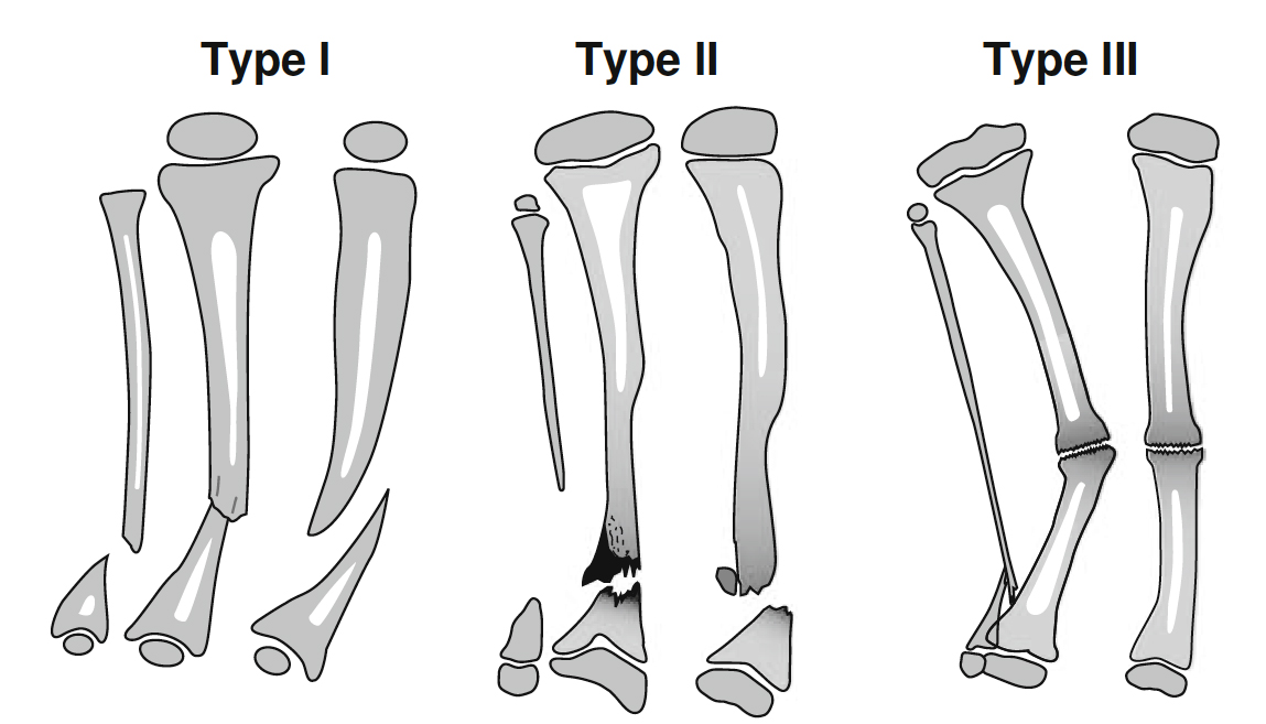
Dr. Paley, in conjunction with Dr. El-Rosassy, developed a classification system designed to indicate prognosis and treatment. The El-Rosassy-Paley classification system divides CPT into three types based on two criteria: the geometry of the bone ends and how mobile they are—that is, whether the bone ends at the pseudarthrosis are thick and stiff or thin and mobile. Another important consideration in the El-Rosassy-Paley classification is whether the patient has undergone a previous, unsuccessful surgery.
Type 1
Atrophic (narrow) bone ends
Mobile pseudarthrosis
No previous surgery
Type 2
Atrophic (narrow) bone ends
Mobile pseudarthrosis
Previous unsuccessful surgery
Type 3
Hypertrophic (wide) bone ends
Stiff pseudarthrosis
Congenital pseudarthrosis of the tibia remains one of the most challenging and misunderstood conditions in orthopedics. The difficulty of treatment lies in the weak healing power at the fracture site, a tendency to refracture after treatment, and the difficulty of stabilizing small osteoporotic bone fragments in small children. Even in cases where a union has been achieved, there is a difficulty in maintaining it. Frequently, the end result is a frustrated child and family who have been through multiple failed surgeries and remain with a limb that is short, deformed, and almost functionless. For this reason, many orthopedic surgeons recommend amputation, particularly after a third failed surgery.
REQUEST AN APPOINTMENT OR BOOK A CONSULANT – Sargam.dange.18@gmail.com
Who gets congenital pseudarthrosis of the tibia?
Congenital pseudarthrosis of the tibia is often associated with neurofibromatosis (NF-1) which has many other serious potential manifestations. However, congenital pseudarthrosis of the tibia is also seen in fibrous dysplasia, osteofibrous dysplasia and cleidocranial dysostosis. A significant percentage remain without a known cause (“idiopathic”).
How is congenital pseudarthrosis of the tibia diagnosed?
Congenital pseudarthrosis of the tibia is typically identified with X-rays and physical examination. Patients who are deemed high risk (e.g., patients with neurofibromatosis) may have screening X-rays performed in early childhood. In some cases, the first sign is a deformity in the leg or a fracture that occurred with minimal trauma.
What will happen during a clinic visit for congenital pseudarthrosis of the tibia?
The doctor will evaluate the patient for neurofibromatosis, a genetic disorder that causes tumors to form on nerve tissue which occurs in 40-50% of patients. The doctor will also ask for a history of previous fractures and examine the leg to determine the range of motion of the false joint. X-rays will be taken to determine the quality of the bone, the condition of the joints, the amount of limb length discrepancy and the severity of any bone abnormalities.
How is congenital pseudarthrosis of the tibia treated?
The primary treatment goal of congenital pseudarthrosis of the tibia is to achieve union of the shin bone (tibia) and to maintain that union. Treatment also addresses the resulting limb length discrepancies and bone deformities. To treat congenital pseudarthrosis of the tibia, the International Center for Limb Lengthening has a combined multimodal, comprehensive treatment program that addresses both the biological and mechanical issues. Many doctors use various elements of our protocol, but we strongly believe that the combined multimodal approach is the most effective.
What happens in surgery for congenital pseudarthrosis of the tibia?
Two to four weeks before surgery, the patient receives a bisphosphonate infusion from our endocrinologist. Bisphosphonates inhibit cells responsible for breaking down bone. It is used to prevent resorption (melting away) of bone graft and to improve local healing response. This can take place in Baltimore or for patients from further away, it can be done closer to home by a local endocrinologist.
In the operating room, the abnormal periosteum is removed from the tibia and fibula (both bones in the lower leg), and the pencil tip ends of the tibia and fibula are slightly trimmed to allow straightening of the leg with stable bone contact to optimize healing.
The tibia is stabilized with intramedullary rods (inside the canal of the bone), and the fibula is stabilized with wires. Newer intramedullary rods have allowed surgeons to avoid crossing and damaging the ankle joint during fixation of almost all cases.
Healthy periosteum and a bone graft are harvested from the pelvis usually on the same side as the tibia pseudarthrosis.
The healthy periosteum, bone graft, and bone morphogenic protein (BMP) are placed in between and around the tibia and fibula bones, generating a large cross-sectional surface area for optimal healing. BMP is a naturally occurring cell signaling molecule that drives bone formation. When used in CPT treatment, it helps increase bone formation in the early stages. This is an off-label use of BMP in the United States (not approved by the FDA for this use). While healing can occur without BMP, it is an important component of the International Center for Limb Lengthening’s multi-faceted approach.
In some cases, an external fixator is applied to the leg to provide rotational stability and to compress the bone ends while they knit. In most cases, an internal plate is used to control the newly realigned limb. In the past, external fixators were a necessity. The combination of the newer ankle-sparing intramedullary rods and improved internal plates gives surgeons another option to maintain improved ankle motion. Your doctor will assess which treatment is best for your child’s unique needs and circumstances.
Once healing has occurred (typically evident by 2 months, and strong by 3-4 months), activity restrictions are lifted, and children can return to normal activities. A plastic leg brace called an ankle foot orthosis (AFO) is worn on the leg under clothing for additional protection.
If an external fixator was used and healing of the tibia has been achieved, then the frame stays on for 3-4 months.
If a proximal lengthening is being performed at the same time, then the frame is in place for 4-6 months. In the external fixation device, the patient is allowed to bear weight as soon as they would like after surgery. After the frame is removed, a long leg cast is placed for 4 weeks. After 4 weeks, the cast is removed and a brace is used for 2-3 months or longer.
If a plate was used instead, the plate is removed 6-12 months after the initial surgery, while the rods remain inside to act as “rebar” to strengthen the newly healed leg and prevent refracture.
After either method is used, regular monitoring of the bone is required on an annual basis. The internal rod may needs to be swapped out every 3-4 years during childhood as the leg grows longer to keep providing protection against refracture.
REQUEST AN APPOINTMENT OR BOOK A CONSULANT – Sargam.dange.18@gmail.com
REQUEST AN APPOINTMENT OR BOOK A CONSULANT – Sargam.dange.18@gmail.com
Congenital diaphragmatic hernia (CDH) occurs when the diaphragm, the muscle that separates the chest from the abdomen, fails to close during prenatal development. This opening allows contents of the abdomen (stomach, intestines and/or liver) to migrate into the chest, impacting the growth and development of the lungs. The lungs will be smaller than expected (pulmonary hypoplasia), and will have less developed blood vessels. This causes high blood pressure in the lungs (pulmonary hypertension).
CDH is one of the most common major congenital anomalies, occurring in 1 of every 2,500 – 3,000 live births. CDH can occur on the left or right side, or rarely on both sides. Newborns affected with CDH will require immediate care at delivery, so early and accurate diagnosis is important.
causes
The cause of CDH is unknown. Typically CDH is an isolated finding although it can occur along with heart disease or a genetic abnormality which can lead to additional complications.
Genetic researchers are using innovative techniques to try to find the potential underlying genetic etiology of CDH. Identifying the genetic cause(s) of CDH will allow our clinicians to better manage and counsel families with an affected child.
Signs and symptoms of CDH
CDH is typically discovered during a routine prenatal ultrasound. The sonographer may notice stomach, intestine, or liver in your baby’s chest where the lungs should be. The baby’s heart may also be pushed to one side by the extra organs in the chest.
diagnosis
CDH is typically detected through a routine ultrasound, which allows doctors to check the position of your baby’s lungs and heart. Following the initial diagnosis, at 22 to 28 weeks of gestation, the SSM Health St. Louis Fetal Care Institute team performs a focused ultrasound, a fetal MRI, and a fetal echocardiogram (echo) to assess the severity of the CDH. We also offer genetic testing to determine if a chromosomal abnormality has caused the CDH. In some situations, additional testing will be required throughout the pregnancy.
The team uses several measurements to understand how the CDH will affect the baby. We measure the lung-to-head ratio (LHR), the liver position, the total lung volume, and the response of the fetal lungs to oxygen. Additionally, we look for any abnormalities that suggest a genetic cause. This critical assessment can often provide insight into the severity of the CDH.
Every case and every outcome is different; however, the liver position and lung-to-head ratio can often be a predictor of the severity of the CDH. “Liver up” means that the liver has migrated through the hole in the diaphragm into the chest cavity, while “liver down” means that it is below the hole in the diaphragm, sitting in the normal position in the abdomen.
REQUEST AN APPOINTMENT OR BOOK A CONSULANT – Sargam.dange.18@gmail.com
What happens at delivery?
Most babies with less severe CDH can be delivered vaginally at full-term, unless there are obstetric indications for cesarean delivery. It is best for these babies to be delivered at a medical center where there is access to a team of pediatric surgeons and neonatologists, along with an established Neonatal Intensive Care Unit (NICU),
At birth, the neonatology team will assess your baby. If necessary, a breathing tube will be inserted and a nasogastric tube will be inserted into the stomach to prevent air from building up in the stomach and intestines. Most babies with CDH will go to SSM Health Cardinal Glennon Children’s Hospital for intensive monitoring prior to surgery.
In severe cases of CDH, we may recommend a special delivery procedure, called an EXIT procedure. If your baby has the signs of a severe CDH, we can discuss the full range of options to care for your baby.
What care is provided after delivery?
The care of babies with CDH can be quite complex, with frequent blood tests, x-rays and machines that are helping to support your baby. Our team will involve and inform our parents about all of the various aspects of their baby’s care. We encourage parents to ask questions to better understand how their baby is responding.
When babies with CDH arrive in the NICU they begin the observation stage of their care until they are ready for hernia repair surgery. In many situations they are kept on a ventilator and given sedation to keep them calm and pain free. Intravenous fluids and nutritional support are also provided. Your baby will not be able to eat until after surgery. If they choose, mothers can pump and store their breast milk until the baby can eat.
Most CDH babies suffer from high blood pressure in the lungs, called pulmonary hypertension. This problem can be quite serious and can delay surgical repair of the diaphragm. Before repair surgery, your baby will be monitored for pulmonary hypertension, and treatments will be started to reduce its severity. For instance, inhaled nitric oxide is often used to help open up the blood vessels in the lungs, decreasing the blood pressure and allowing for better oxygen delivery.
CDH babies typically undergo surgery to repair the hole in their diaphragm once pulmonary hypertension has subsided. The CDH team will work together with you to develop the optimal care plan for you and your baby.
During surgery, the surgeons will assess the size and location of the defect to determine which type of closure is required. During the CDH repair procedure a small incision in made under the rib cage to give the surgeon access to the diaphragm and misplaced organs. The surgeon will gently reposition the organs in the abdomen, and close the hole in the diaphragm. Smaller hernias may be repaired with stitches, but larger defects require a medical patch.
After surgery, your baby will return to the NICU to recover and heal. The stress of the surgery may cause your baby’s condition to initially get worse before it gets better, and treatment may require additional sedation, ventilation, blood pressure medications or ECMO.
The amount of time spent in the NICU after surgery varies from patient to patient depending on the severity of their case. The CDH team will work to wean your baby off the ventilator and pain medications as quickly as possible, and to get them eating and gaining weigh
Management of CDH during pregnancy
After all testing is complete, our team, led by a maternal-fetal medicine specialist and a pediatric surgeon both with experience managing pregnancies affected by CDH, meets with you and your family. Together we review imaging and test results, discuss the diagnosis, explain treatment options and potential outcomes, and answer any questions you may have.
You will typically be seen every four weeks for follow up until later in pregnancy when you’ll be seen more frequently by an obstetrical team experienced in managing pregnancies affected by CDH and led by a maternal-fetal medicine specialist. During your follow up ultrasounds the lung measurements will be updated, your baby’s growth will be carefully watched, and the amount of amniotic fluid around the baby will be measured. Weekly testing begins at 34 weeks to closely monitor fetal well-being.
At this time, relocation is necessary if you live more than one hour away from our center. You will need to relocate sooner if you undergo prenatal intervention, if extra fluid develops around your baby (polyhydramnios), or if there are signs of preterm labor. This will ensure that you are nearby in the event labor occurs or delivery is indicated earlier than expected. Our social workers will help coordinate housing options based upon your family’s needs.
REQUEST AN APPOINTMENT OR BOOK A CONSULANT – Sargam.dange.18@gmail.com
REQUEST AN APPOINTMENT OR BOOK A CONSULANT – Sargam.dange.18@gmail.com
Infectious arthritis is an infection in a joint. It may also be referred to as septic arthritis. It occurs when an infection caused by a bacteria or virus spreads to a joint or the fluid surrounding the joint. This fluid is called the synovial fluid. This infection usually begins in another area of the body and spreads through the bloodstream to the joint tissue. The infection may also enter the body through surgery, open wounds, or injections.
Infectious arthritis usually only occurs in one joint. The condition typically affects a large joint such as the knee, hip, or shoulder. It occurs more often in children, older adults, and people who use illegal drugs.
Septic arthritis is a painful infection in a joint that can come from germs that travel through your bloodstream from another part of your body. Septic arthritis can also occur when a penetrating injury, such as an animal bite or trauma, delivers germs directly into the joint.
Infants and older adults are most likely to develop septic arthritis. People who have artificial joints are also at risk of septic arthritis. Knees are most commonly affected, but septic arthritis also can affect hips, shoulders and other joints. The infection can quickly and severely damage the cartilage and bone within the joint, so prompt treatment is crucial.
Treatment involves draining the joint with a needle or during surgery. Antibiotics also are usually needed.
Causes
Septic arthritis can be caused by bacterial, viral or fungal infections. Bacterial infection with Staphylococcus aureus (staph) is the most common cause. Staph commonly lives on even healthy skin.
Septic arthritis can develop when an infection, such as a skin infection or urinary tract infection, spreads through your bloodstream to a joint. Less commonly, a puncture wound, drug injection, or surgery in or near a joint — including joint replacement surgery — can give the germs entry into the joint space.
The lining of your joints has little ability to protect itself from infection. Your body’s reaction to the infection — including inflammation that can increase pressure and reduce blood flow within the joint — contributes to the damage.
Symptoms
Septic arthritis typically causes extreme discomfort and difficulty using the affected joint. The joint could be swollen, red and warm, and you might have a fever.
If septic arthritis occurs in an artificial joint (prosthetic joint infection), signs and symptoms such as minor pain and swelling may develop months or years after knee replacement or hip replacement surgery. Also, a loosening of the joint may occur, which causes pain while moving the joint or while putting weight on the joint. Typically, the pain goes away when at rest. In extreme cases, the joint may become dislocated.
The symptoms of infectious arthritis can vary depending on your age and the medications you’re taking. The symptoms may include:
severe pain that worsens with movement
swelling of the joint
warmth and redness around the joint
a fever
chills
fatigue
weakness
decreased appetite
a rapid heart rate
irritability
Diagnosis
Your doctor will examine your joint and ask you questions about your symptoms. If they suspect you have infectious arthritis, they may order additional tests.
An arthrocentesis is a test frequently used to diagnose this condition. It involves inserting a needle into the affected joint to take a sample of synovial fluid. The sample is sent to the lab to be examined for color, consistency, and the presence of white blood cells and bacteria. The information from this test can tell your doctor if you have an infection in the joint and what is causing the infection.
Your doctor may also take a blood sample from you. This is another way to check your white blood cell count and to determine if any bacteria are present in your bloodstream. This information can help your doctor determine the severity of the infection.
Imaging tests may also be ordered to confirm the presence of infection. These tests can also help your doctor see if your joint has been damaged by the infection. Imaging tests used for infectious arthritis include:
X-rays
MRI scans
CT scans
nuclear scans
REQUEST AN APPOINTMENT OR BOOK A CONSULANT – Sargam.dange.18@gmail.com
Risk factors
Risk factors for septic arthritis include:
Existing joint problems. Chronic diseases and conditions that affect your joints — such as osteoarthritis, gout, rheumatoid arthritis or lupus — can increase your risk of septic arthritis, as can previous joint surgery and joint injury.
Having an artificial joint. Bacteria can be introduced during joint replacement surgery, or an artificial joint may become infected if germs travel to the joint from a different area of the body through the bloodstream.
Taking medications for rheumatoid arthritis. People with rheumatoid arthritis have a further increase in risk because of medications they take that can suppress the immune system, making infections more likely to occur. Diagnosing septic arthritis in people with rheumatoid arthritis is difficult because many of the signs and symptoms are similar.
Skin fragility. Skin that breaks easily and heals poorly can give bacteria access to your body. Skin conditions such as psoriasis and eczema increase your risk of septic arthritis, as do infected skin wounds. People who regularly inject drugs also have a higher risk of infection at the site of injection.
Weak immune system. People with a weak immune system are at greater risk of septic arthritis. This includes people with diabetes, kidney and liver problems, and those taking drugs that suppress their immune systems.
Joint trauma. Animal bites, puncture wounds or cuts over a joint can put you at risk of septic arthritis.
Having a combination of risk factors puts you at greater risk than having just one risk factor does.
When to see a doctor
See your doctor if you have severe pain in a joint that comes on suddenly. Prompt treatment can help minimize joint damage.
If you have an artificial joint, see your doctor if you experience pain while using the joint.
Complications
If treatment is delayed, septic arthritis can lead to joint degeneration and permanent damage. If septic arthritis affects an artificial joint, complications may include joint loosening or dislocation.
Treatment
Prescription Drugs
Treatment for infectious arthritis caused by a bacteria usually begins with antibiotics to kill the bacteria causing the infection. Your doctor will use the information from your tests to choose an antibiotic that’s effective for the type of bacteria present in your joint. The infection needs to be treated promptly and aggressively to prevent osteoarthritis and damage to your joint. As a result, your doctor may order intravenous antibiotics, which are given through your veins. This treats the infection more quickly than oral antibiotics. Most people begin to feel better within 48 hours of their first antibiotic treatment.
Your doctor may also prescribe oral antibiotics to treat the infection. Oral antibiotics for infectious arthritis usually need to be taken for six to eight weeks. It’s important to take the entire course of antibiotics to treat the infection effectively.
Your doctor will prescribe antifungal medication instead of antibiotics if a fungus is causing your infection.
Infectious arthritis caused by a virus doesn’t require medication.
REQUEST AN APPOINTMENT OR BOOK A CONSULANT – Sargam.dange.18@gmail.com
Synovial Fluid Drainage
Many people with infectious arthritis need to have their synovial fluid drained. This is done to remove the infected fluid, ease pain and swelling, and prevent further damage to the joint. Synovial fluid is often drained using arthroscopy, but it can be done in an open surgical procedure.
With arthroscopy, your doctor will make several small incisions near the affected joint. Then, they’ll insert a small tube containing a camera into the incision. Your doctor will use the camera image to guide them in suctioning the infected fluid from your joint. Usually, a drain or tube will be inserted and left in the joint to keep the joint from swelling again. This drain is then removed in a few days.
Sometimes, a doctor can use a small needle to remove infected fluid without requiring surgery. This is called arthrocentesis. This procedure often has to be repeated over the course of several days to ensure the fluid has been removed.
Other Treatment Options
Most cases of infectious arthritis require surgery, such as arthroscopy or an open procedure, to wash out the joint. On occasion, surgery is required to remove any damaged sections of the joint or replace the joint, but this is only done after the infection has been treated.
Other treatment methods to reduce pain may be used along with treatment for the infection. These methods include:
using nonsteroidal anti-inflammatory drugs
resting the joint
splinting the affected joint
going to physical therapy
Physical Therapy Management
Patients with septic arthritis are usually managed initially in the inpatient hospital setting and require an interprofessional team of caregivers including the primary care provider, physical and occupational therapist, nurses and wound care team. At the time of discharge, some patients can return home while others with increased debility may need ongoing physical therapy at either an acute or subacute rehabilitation facility. The social workers in the hospital help to ensure a smooth transition from inpatient to outpatient care..
The initial treatment of infectious arthritis is outside of the scope of physical therapy. It is important, first, for the physical therapist to recognize the signs and symptoms of the infection and refer out for other medical treatment. Subjective history, in combination with the physical therapist’s objective findings, is important in order to recognize the risk factors that make septic arthritis the likely diagnosis. It is important to immobilize the joint in this stage to best manage the patient’s pain and to decrease the likelihood of doing further damage to the joint until proper treatment can occur.
Once the patient receives a round of antibiotic treatment in combination with either joint aspiration, debridement, or arthroscopy, the patient may then be referred back to physical therapy including
educate on how to properly protect the affected joint.
Gentle mobilization of the infected joint can begin if the patient is responding well following 5 days of medical treatment.
Once the infection is well-managed, current evidence states the patient will usually respond best to aggressive physical therapy to allow maximum post-infection functioning.
Physical therapy needs to consist of allowing the joint to be in its functional position and positioning the joint to allow passive range of motion activities.
REQUEST AN APPOINTMENT OR BOOK A CONSULANT – Sargam.dange.18@gmail.com
REQUEST AN APPOINTMENT OR BOOK A CONSULANT – Sargam.dange.18@gmail.com
Tuberculous synovitis frequently presents as a monoarthritis of weight-bearing joints such as the hip, knee, or ankle. Owing to its low incidence in developed countries, the diagnosis is often delayed for months to years. Early diagnosis with a synovial biopsy permits prompt antituberculous therapy and substantially improves the prospect of preservation of joint structure and function. Initial treatment typically includes combination therapy with four drugs (isoniazid, rifampin, pyrazinamide, and streptomycin or ethambutol) because of the frequency of isoniazid resistance. Antimicrobial therapy should be of at least 9 months’ duration, longer in immunocompromised hosts. Partial synovectomy and other surgical procedures should be restricted to joints with severe cartilage destruction, large abscesses, joint deformity, multiple drug resistance, or atypical mycobacteria.
Tubercular bacilli reach the joint space via the blood stream through subsynovial vessels, or indirectly from epiphyseal (more common in adults) or metaphyseal (more common in children) lesions, which erode into the joint space. Articular cartilage destruction begins peripherally and the weightbearing surfaces are preserved for a few months, providing the potential for good functional recovery with effective treatment in patients with early disease.
The disease may start in bone or in the synovial membrane, but one rapidly infects the other. The initial focus starts in the metaphysis in childhood or at the end of the bone in adults. An example of articular tuberculosis of the typical osseous areas of predilection for hip disease.
Osseous Changes and Tubercular Sequestra
After the infection, marked hyperemia and severe osteoporosis occur. The softened bone easily yields under the effect of gravity and muscle action, leading to compression, collapse, or deformation. Necrosis also may be caused by ischemic infarction of segments of bone.
Sequestration gives the appearance of coarse sand and rarely produces a radiologically visible sequestrum. Because of loss of nutrition, the adjacent articular cartilage or the intervening disc degenerate and also may become separated as sequestra. Some of the radiologically visible smaller sequestra in tuberculous cavities may result from calcification of caseous matter.
The Future Course of Tubercle
Before the availability of antitubercular drugs, the 5–year followup mortality of patients with osteoarticular tuberculosis was approximately 30.Modern antitubercular agents have changed the outlook considerably. Depending on the sensitivity pattern, host resistance, and the stage of the lesion at the inception of treatment, the tuberculous lesion may behave as follows. It may resolve completely; the disease may heal with residual deformity and loss of function; the lesion may be walled off completely and the caseous tissue may calcify; low-grade chronic fibromatous granulating and caseating lesion may persist (grumbling disease); and the infection may spread locally by contiguity and systematically by the blood stream
DIAGNOSIS OF TB INFECTION
Methods
Tuberculin Skin Test (TST)
Interferon Gamma Release Assays (IGRAs)
Enzyme-Linked Immunosorbent Assays (ELISA)
Bacteriology
Radiology
Computed Tomography (CT)
Magnetic Resonance Imaging (MRI)
Polymerase Chain Reaction (PCR)
Synovial fluid examination
Synovial biopsy
Blood
A relative lymphocytosis, low hemoglobin, and increased erythrocyte sedimentation rate often are found in patients with the active stage of disease. An increased erythrocyte sedimentation rate, however, is not necessarily proof of activity of the infection. Its repeated estimation at 3- to 6-month intervals gives an index of the activity of the disease.
Mantoux Test
As a rule, a positive reaction is present in a patient infected with tuberculosis for more than 1 month. A negative test, in general, rules out the disease. The tuberculin test rarely may be negative although active tuberculosis is present, such as in immune deficiency states.
Biopsy
Whenever there is doubt (particularly in the early stages) it is mandatory to prove the diagnosis by obtaining a biopsy specimen of the diseased tissue (granulations, synovium, bone, lymph nodes, or margins of tuberculous ulcers). Microscopic examination of an aspiration, core biopsy, needle biopsy, or open biopsy will reveal typical tubercles in patients who are not treated. Epithelioid cells surrounded by lymphocytes, even without central necrosis or peripheral foreign-body giant cells, are adequate histologic evidence of tuberculosis in a patient who is suspected to have the disease. At the time of open biopsy of a joint or bone, the orthopaedic surgeon should do therapeutic synovectomy or curettage. The infections of bone and joint that present as granulomatous lesions in order of frequency are tuberculosis, mycotic infection, brucellosis, sarcoidosis, and tuberculoid leprosy.
Guinea Pig Inoculation
The tuberculous pus, joint aspirate, or diseased material may be injected intraperitoneally into a guinea pig. Examination in positive cases shows tubercles on the peritoneum 5 to 8 weeks later. Although it currently is not considered a cost-effective test, it perhaps is the most reliable proof of tuberculosis.
Smear, Culture, and Serology
The material prepared for guinea pig inoculation also may be submitted for smear and culture examination for acid-fast bacilli. In superficial joints, one may be able to aspirate synovial fluid. Analysis of synovial fluid does not provide pathognomonic information; however, in general the leukocyte count is elevated to approximately 20,000 mm3, there is a lowered glucose level, and poor mucin. Clear synovial aspirate is not an appropriate material for microbiologic investigation; however, it is an excellent material for polymerase chain reaction and nucleic acid probes.
REQUEST AN APPOINTMENT OR BOOK A CONSULANT – Sargam.dange.18@gmail.com
Principles of Treatment of Osteoarticular Tuberculosis
Modern drugs promote the healing of sinuses, ulcers, and abscesses in patients previously unresponsive to extensive surgery. They also eliminate the danger of postoperative miliary and meningeal disease caused by dissemination of the tuberculous infection.
Death caused by uncontrolled disease, meningitis, miliary tuberculosis, amyloidosis, paralysis, and crippling now is rare. If a patient is diagnosed early and treated vigorously, healing can be accomplished without residual joint ankylosis or deformity.
With the use of modern drugs, the indications for surgery have become universally more selective and directed toward the prevention and correction of deformities, and the improvement in function of the diseased joints. At the stage of tuberculous arthritis, if abscess formation has not occurred, the natural outcome generally is a fibrous ankylosis. If an abscess discharges and sinuses develop, the outcome may be a bony ankylosis. The prognosis in articular tuberculosis depends on the stage of the disease. when the specific treatment is started. Concomitant disease must be treated and hospitalization is necessary only for patients with complications, or for patients requiring traction under supervision to correct deformities.
Category of treatment
Category of TB cases
Anti-TB drug regimens
Intensive phase
Continuation phase
I
New Patient Regimen New smear-positive PTB Smear-negative PTB with extensive Parenchymal involvement Severe forms of EPTB other than TB meningitis
2HRZE
4HR
II
New Patient Regimen Smear-negative PTB without extensive P Parenchymal involvement Less severe forms of EPTB (e.g., TB cervical adenitis)
2HRZ
4HR
III
New Patient Regimen TB meningitis
2HRZSa
4HR
IV
Retreatment regimen Previously treated smear-positive PTB (relapse, treatment after interruption or treatment failure) If low risk for MDR-TB or risk unknown, continue with retreatment regimen If high risk for MDR-TB, use MDR-TB regimen below
2HRZES/1HRZE
5HRE
V
MDR Regimen MDR-TB
Individualized regimens
Table 2: Treatment regimens for tuberculosis recommended by the WHO . Other regimens are recommended for the treatment of TB meningitis, including replacing streptomycin with ethionamide and treating for 9-12 months. E: Ethambutol; EPTB: Extra-Pulmonary Tuberculosis; H: Isoniazid; HIV: Human Immunodeficiency Virus; MDR-TB: Multi-Drug Resistant Tuberculosis; PTB: Pulmonary Tuberculosis; R: Rifampicin; S: Streptomycin; Z: Pyrazinamide; 2HRZ 4HR: Denotes a Two-month Intensive Phase of Daily Isoniazid, Rifampicin, and Pyrazinamide followed by a Four-month Continuation Phase of Daily Isoniazid and Rifampicin
Rest, Immobilization, and Braces
In the active stage of disease, the joints are rested in the position of function using removable splints. Prolonged immobilization can lead to spontaneous ankylosis when joints are grossly destroyed. Patients with early disease are allowed 1 to 2 hours of intermittent, guarded active and assisted exercises while taking antitubercular drugs, with the aim of retaining a useful range of movement in the functional arc of the involved joint. Traction helps to correct deformity and to rest the diseased part. Gradual ambulation is encouraged with the help of suitable braces approximately 3 months after the start of treatment while healing is progressing. As the disease heals and pain subsides, weightbearing and activity are permitted. If there is steady progress, activity is increased within the limits of discomfort. The use of a brace is discontinued gradually after approximately 2 years.
Treatment of Abscess, Effusion, and Sinus
Palpable and large joint effusions are aspirated and 1000 mg of streptomycin alone or combined with injectable isoniazid (300 mg) is instilled at each aspiration. Local concentrations of antibiotics after parenteral administration may make this local instillation unnecessary. Open drainage of an abscess is indicated if aspiration fails. Radiologically visible paravertebral abscess shadows do not need to be drained, unless decompression is done in patients with paraplegia or when diseased vertebrae are debrided. A prevertebral abscess in the cervical region is drained if it causes difficulty in swallowing or breathing. Drainage of a large paravertebral abscess also may be considered when its radiologic size increases markedly despite treatment.
A majority of ulcers and sinuses heal within 6 to 12 weeks under the influence of systemic antitubercular drugs. Less than 1% of patients with sinuses require longer treatment and excision of the tract, with or without debridement. Sinus ramification always is greater than can be appreciated and complete surgical excision therefore is impractical.
REQUEST AN APPOINTMENT OR BOOK A CONSULANT – Sargam.dange.18@gmail.com
Antitubercular Chemotherapy
Combination chemotherapy should be used for an adequate length of time. Most of the antitubercular drugs potentially are toxic and resistance or intolerance to the drugs should be suspected when a patient fails to respond.
Multidrug Resistant Tuberculosis and Patients Who do not Respond
If the disease is caused by organisms resistant to isoniazid and rifampicin (multidrug resistant tuberculosis), if the disease is not controlled within 4 to 5 months, or if despite multidrug therapy, more active tuberculous lesions appear, one has to resort to second line drugs or potential antituberculous drugs. The situation is desperate in patients who do not respond. Immunomodulation in conjunction with drugs may be used in such patients. A favorable response was reported in approximately 85% of patients.
Pending the availability of better immunomodulation techniques.the current author has evolved the following outline during the past 12 years to upgrade cell-mediated immunity. In brief, 150 mg of levamisol is given at night for 3 days at weekly intervals for a total of 45 tablets. Four injections are administered once a month. The first and second infections are 0.1 mL intradermal (Bacillus Calmett-Guérin) injections and the third and fourth are intramuscular DPT injections (diphtheria vaccine + tetanus vaccine + Bordetella pertussis 20,000 million per 0.5 mL).
Surgery in Patients with Tuberculosis of Bones and Joints
No surgery is a substitute for a prolonged course of antitubercular drugs. A trial of conservative treatment is justified in most patients before surgery is contemplated. Nonoperative treatment usually is adequate in patients with pure synovial tuberculosis, low-grade or early arthritis of any joint, and even advanced (Stage III or IV) arthritis, especially in the upper extremity.
Surgery only should be considered once the general condition of the patient is stabilized by drug therapy, before the development of drug resistance. In general, a minimum of 1 to 4 weeks of therapy is advisable before any major surgical intervention.
Relapse of Osteoarticular Tuberculosis or Recurrence of Complications
The incidence of relapse or recurrence is unknown because these complications may occur at any period during the lifetime of a patient, whether the initial treatment included excisional surgery. Reactivation may occur in 2% to 5% of patients as late as 20 years or more after apparent healing.
The causes of reactivation include prolonged use of systemic cortisone therapy, malnutrition, the development of diabetes or an immune deficiency state, or a surgical procedure or injury to the previously infected area. Dormant bacilli persisting in tissue for years may start multiplying under such circumstances.
Tuberculosis of the Spine
Vertebral tuberculosis accounts for 50% of all cases of skeletal tuberculosis. In the majority of patients, the disease typically started in the paradiscal region. Narrowing of the disc often is the earliest radiologic finding. Any reduction in disc space, if it is associated with a loss of definition of the paradiscal margins of the vertebrae, suggests tuberculosis and occurs before the appearance of frank osseous destruction. These changes usually are evident only after infection has been present for 3 to 6 months, although advanced imaging may detect changes at approximately 6 weeks.
Most cases of tuberculosis of the spine heal without surgical intervention. However, uncertainty about the diagnosis, progressive bone destruction, and neurologic symptoms despite chemotherapy are definite indications for surgery in the active stage of disease.
REQUEST AN APPOINTMENT OR BOOK A CONSULANT – Sargam.dange.18@gmail.com