REQUEST AN APPOINTMENT OR BOOK A CONSULANT – Sargam.dange.18@gmail.com
Precocious puberty is when a child’s body begins changing into that of an adult (puberty) too soon. When puberty begins before age 8 in girls and before age 9 in boys, it is considered precocious puberty.
Puberty includes rapid growth of bones and muscles, changes in body shape and size, and development of the body’s ability to reproduce.
The cause of precocious puberty often can’t be found. Rarely, certain conditions, such as infections, hormone disorders, tumors, brain abnormalities or injuries, may cause precocious puberty. Treatment for precocious puberty typically includes medication to delay further development.
Symptoms
Precocious puberty signs and symptoms include development of the following before age 8 in girls and before age 9 in boys.
Breast growth and first period in girls
Enlarged testicles and penis, facial hair and deepening voice in boys
Pubic or underarm hair
Rapid growth
Acne
Adult body odor
When to see a doctor
Make an appointment with your child’s doctor for an evaluation if your child has any of the signs or symptoms of precocious puberty.
Causes
To understand what causes precocious puberty in some children, it’s helpful to know what causes puberty to begin. The brain starts the process with the production of a hormone called gonadotropin-releasing hormone (GnRH).
When this hormone reaches the pituitary gland — a small bean-shaped gland at the base of your brain — it leads to the production of more hormones in the ovaries for females (estrogen) and the testicles for males (testosterone).
Estrogen is involved in the growth and development of female sexual characteristics. Testosterone is responsible for the growth and development of male sexual characteristics.
Why this process begins early in some children depends on whether they have central precocious puberty or peripheral precocious puberty.
Central precocious puberty
The cause for this type of precocious puberty often can’t be identified.
In central precocious puberty, the puberty process starts too soon. The pattern and timing of the steps in the process are otherwise normal. For the majority of children with this condition, there’s no underlying medical problem and no identifiable reason for the early puberty.
In rare cases, central precocious puberty may be caused by:
A tumor in the brain or spinal cord (central nervous system)
A defect in the brain present at birth, such as excess fluid buildup (hydrocephalus) or a noncancerous tumor (hamartoma)
Radiation to the brain or spinal cord
Injury to the brain or spinal cord
McCune-Albright syndrome — a rare genetic disease that affects bones and skin color and causes hormonal problems
Congenital adrenal hyperplasia — a group of genetic disorders involving abnormal hormone production by the adrenal glands
Hypothyroidism — a condition in which the thyroid gland doesn’t produce enough hormones
REQUEST AN APPOINTMENT OR BOOK A CONSULANT – Sargam.dange.18@gmail.com
Peripheral precocious puberty
Estrogen or testosterone in your child’s body causes this type of precocious puberty.
The less common peripheral precocious puberty occurs without the involvement of the hormone in your brain (GnRH) that normally triggers the start of puberty. Instead, the cause is release of estrogen or testosterone into the body because of problems with the ovaries, testicles, adrenal glands or pituitary gland.
In both girls and boys, the following may lead to peripheral precocious puberty:
A tumor in the adrenal glands or in the pituitary gland that releases estrogen or testosterone
McCune-Albright syndrome, a rare genetic disorder that affects the skin color and bones and causes hormonal problems
Exposure to external sources of estrogen or testosterone, such as creams or ointments
In girls, peripheral precocious puberty may also be associated with:
Ovarian cysts
Ovarian tumors
In boys, peripheral precocious puberty may also be caused by:
A tumor in the cells that make sperm (germ cells) or in the cells that make testosterone (Leydig cells).
A rare disorder called gonadotropin-independent familial sexual precocity, which is caused by a defect in a gene, can result in the early production of testosterone in boys, usually between ages 1 and 4.
Risk factors
Factors that increase a child’s risk of precocious puberty include:
Being a girl. Girls are much more likely to develop precocious puberty.
Being African-American. Precocious puberty appears to affect African-Americans more often than children of other races.
Being obese. Children who are significantly overweight have a higher risk of developing precocious puberty.
Being exposed to sex hormones. Coming in contact with an estrogen or testosterone cream or ointment, or other substances that contain these hormones (such as an adult’s medication or dietary supplements), can increase your child’s risk of developing precocious puberty.
Having other medical conditions. Precocious puberty may be a complication of McCune-Albright syndrome or congenital adrenal hyperplasia — conditions that involve abnormal production of the male hormones (androgens). In rare cases, precocious puberty may also be associated with hypothyroidism.
Having received radiation therapy of the central nervous system. Radiation treatment for tumors, leukemia or other conditions can increase the risk of precocious puberty.
Complications
Possible complications of precocious puberty include:
Short height. Children with precocious puberty may grow quickly at first and be tall, compared with their peers. But, because their bones mature more quickly than normal, they often stop growing earlier than usual. This can cause them to be shorter than average as adults. Early treatment of precocious puberty, especially when it occurs in very young children, can help them grow taller than they would without treatment.
Social and emotional problems. Girls and boys who begin puberty long before their peers may be extremely self-conscious about the changes occurring in their bodies. This may affect self-esteem and increase the risk of depression or substance abuse.
Prevention
Some of the risk factors for precocious puberty, such as sex and race, can’t be avoided. But, there are things you can do to reduce your child’s chances of developing precocious puberty, including:
Keeping your child away from external sources of estrogen and testosterone — such as prescription medications for adults in the house or dietary supplements containing estrogen or testosterone
Encouraging your child to maintain a healthy weight
REQUEST AN APPOINTMENT OR BOOK A CONSULANT – Sargam.dange.18@gmail.com
REQUEST AN APPOINTMENT OR BOOK A CONSULANT – Sargam.dange.18@gmail.com
Rhesus (Rh) factor is an inherited protein found on the surface of red blood cells. If your blood has the protein, you’re Rh positive. If your blood lacks the protein, you’re Rh negative.
Rh positive is the most common blood type. Having an Rh negative blood type is not an illness and usually does not affect your health. However, it can affect your pregnancy. Your pregnancy needs special care if you’re Rh negative and your baby is Rh positive (Rh incompatibility). A baby can inherit the Rh factor from either parent.
Your health care provider will recommend a blood type and Rh factor screening test during your first prenatal visit. This will identify whether your blood cells carry the Rh factor protein.
What’s an Rh Factor?
An Rh factor is a protein found on some red blood cells (RBCs). Not everyone carries this protein, though most do. They are Rh-positive. People who don’t carry the protein are Rh-negative.
What if Parents Don’t Have the Same Rh Factor?
When a mother-to-be and father-to-be are not both positive or negative for Rh factor, it’s called Rh incompatibility.
For example:
If a woman who is Rh negative and a man who is Rh positive conceive a baby, the fetus may have Rh-positive blood, inherited from the father. (About half of the children born to an Rh-negative mother and Rh-positive father will be Rh-positive.)
Rh incompatibility usually isn’t a problem if it’s the mother’s first pregnancy. That’s because the baby’s blood does not normally enter the mother’s circulatory system during the pregnancy.
During the birth, though, the mother’s and baby’s blood can mix. If this happens, the mother’s body recognizes the Rh protein as a foreign substance. It then might begin making antibodies (proteins that act as protectors if foreign cells enter the body) against the Rh protein.
Rh-negative pregnant women can be exposed to the Rh protein that might cause antibody production in other ways too. These include:
blood transfusions with Rh-positive blood
miscarriage
ectopic pregnancy
Why it’s done
During pregnancy, problems can occur if you’re Rh negative and the baby you’re carrying is Rh positive. Usually, your blood doesn’t mix with your baby’s blood during pregnancy. However, a small amount of your baby’s blood could come in contact with your blood during delivery or if you experience bleeding or abdominal trauma during pregnancy. If you’re Rh negative and your baby is Rh positive, your body might produce proteins called Rh antibodies after exposure to the baby’s red blood cells.
The antibodies produced aren’t a problem during the first pregnancy. The concern is with your next pregnancy. If your next baby is Rh positive, these Rh antibodies can cross the placenta and damage the baby’s red blood cells. This could lead to life-threatening anemia, a condition in which red blood cells are destroyed faster than the baby’s body can replace them. Red blood cells are needed to carry oxygen throughout the body.
If you’re Rh negative, you might need to have another blood test — an antibody screen — during your first trimester, during week 28 of pregnancy and at delivery. The antibody screen is used to detect antibodies to Rh positive blood. If you haven’t started to produce Rh antibodies, you’ll likely need an injection of a blood product called Rh immune globulin. The immune globulin prevents your body from producing Rh antibodies during your pregnancy.
If your baby is born Rh negative, no additional treatment is needed. If your baby is born Rh positive, you’ll need another injection shortly after delivery.
If you’re Rh negative and your baby might be or is Rh positive, your health care provider might recommend an Rh immune globulin injection after situations in which your blood could come into contact with the baby’s blood, including:
Miscarriage
Abortion
Ectopic pregnancy — when a fertilized eggs implants somewhere outside the uterus, usually in a fallopian tube
Removal of a molar pregnancy — a noncancerous (benign) tumor that develops in the uterus
Amniocentesis — a prenatal test in which a sample of the fluid that surrounds and protects a baby in the uterus (amniotic fluid) is removed for testing or treatment
Chorionic villus sampling — a prenatal test in which a sample of the wispy projections that make up most of the placenta (chorionic villi) is removed for testing
Cordocentesis — a diagnostic prenatal test in which a sample of the baby’s blood is removed from the umbilical cord for testing
Bleeding during pregnancy
Abdominal trauma during pregnancy
The external manual rotation of a baby in a breech position — such as buttocks first — before labor
Delivery
If the antibody screen shows that you’re already producing antibodies, an injection of Rh immune globulin won’t help. Your baby will be carefully monitored. He or she might be given a blood transfusion through the umbilical cord during the pregnancy or immediately after delivery if necessary.
REQUEST AN APPOINTMENT OR BOOK A CONSULANT – Sargam.dange.18@gmail.com
Mother’s Rh factor
Father’s Rh factor
Baby’s Rh factor
Precautions
Rh positive
Rh positive
Rh positive
None
Rh negative
Rh negative
Rh negative
None
Rh positive
Rh negative
Could be Rh positive or Rh negative
None
Rh negative
Rh positive
Could be Rh positive or Rh negative
Rh immune globulin injections
What you can expect
An Rh factor test is a basic blood test. The blood sample is usually taken during the first prenatal visit and sent to a lab for analysis. No special preparation is necessary.
Results
If you’re Rh positive, no action is needed.
If you’re Rh negative and your baby is Rh positive, there’s a potential for your body to produce antibodies that could be harmful during a subsequent pregnancy. If you have vaginal bleeding at any time during pregnancy, contact your health care provider immediately. Also, talk with your health care provider about scheduling an Rh immune globulin injection during your pregnancy and remind your health care team of your Rh status during labor.
When Is a Baby at Risk?
Rh antibodies are harmless until the mother’s second or later pregnancies. If she is ever carrying another Rh-positive child, her Rh antibodies will recognize the Rh proteins on the surface of the baby’s blood cells as foreign. Her antibodies will pass into the baby’s bloodstream and attack those cells.
This can make the baby’s red blood cells swell and rupture. This is known as hemolytic or Rh disease of the newborn. It can make a baby’s blood count get very low.
How Is Rh Incompatibility Treated?
If a pregnant woman has the potential to develop Rh incompatibility, doctors give her a series of two Rh immune-globulin shots during her first pregnancy. She’ll get:
the first shot around the 28th week of pregnancy
the second shot within 72 hours of giving birth
Rh immune-globulin acts like a vaccine. It prevents the mother’s body from making any Rh antibodies that could cause serious health problems in the newborn or affect a future pregnancy.
A woman also might get a dose of Rh immune-globulin if she has a miscarriage, an amniocentesis, or any bleeding during pregnancy.
If a doctor finds that a woman has already developed Rh antibodies, her pregnancy will be closely watched to make sure that those levels are not too high.
In rare cases, if the incompatibility is severe and a baby is in danger, the baby can get special blood transfusions called exchange transfusions either before birth (intrauterine fetal transfusions) or after delivery. Exchange transfusions replace the baby’s blood with blood with Rh-negative blood cells. This stabilizes the level of red blood cells and minimizes damage from Rh antibodies already in the baby’s bloodstream.
REQUEST AN APPOINTMENT OR BOOK A CONSULANT – Sargam.dange.18@gmail.com
REQUEST AN APPOINTMENT OR BOOK A CONSULANT – Sargam.dange.18@gmail.com
Menopause is the time that marks the end of your menstrual cycles. It’s diagnosed after you’ve gone 12 months without a menstrual period. Menopause can happen in your 40s or 50s, but the average age is 51 in the United States.
Menopause is a natural biological process. But the physical symptoms, such as hot flashes, and emotional symptoms of menopause may disrupt your sleep, lower your energy or affect emotional health. There are many effective treatments available, from lifestyle adjustments to hormone therapy.
Menopause occurs when a woman hasn’t menstruated in 12 consecutive months and can no longer become pregnant naturally. It usually begins between the ages of 45 and 55, but can develop before or after this age range.
Menopause can cause uncomfortable symptoms, such as hot flashes and weight gain. For most women, medical treatment isn’t needed for menopause.
Read on to learn what you need to know about menopause.
When does menopause begin and how long does it last?
Most women first begin developing menopause symptoms about four years before their last period. Symptoms often continue until about four years after a woman’s last period.
A small number of women experience menopause symptoms for up to a decade before menopause actually occurs, and 1 in 10 women experience menopausal symptoms for 12 years following their last period.
The median age for menopause is 51, though it may occur on average up to two years earlier for Black and Latina women. More studies are needed to understand the onset of menopause for women of color.
There are many factors that help determine when you’ll begin menopause, including genetics and ovary health. Perimenopause occurs before menopause. Perimenopause is a time when your hormones begin to change in preparation for menopause.
It can last anywhere from a few months to several years. Many women begin perimenopause some point after their mid-40s. Other women skip perimenopause and enter menopause suddenly.
About 1 percent of women begin menopause before the age of 40, which is called premature menopause or primary ovarian insufficiency. About 5 percent of women undergo menopause between the ages of 40 and 45. This is referred to as early menopause.
Perimenopause vs. menopause vs. postmenopause
During perimenopause, menstrual periods become irregular. Your periods may be late, or you may completely skip one or more periods. Menstrual flow may also become heavier or lighter.
Menopause is defined as a lack of menstruation for one full year.
Postmenopause refers to the years after menopause has occurred.
What are the symptoms of menopause?
Every woman’s menopause experience is unique. Symptoms are usually more severe when menopause occurs suddenly or over a shorter period of time.
Conditions that impact the health of the ovary, like cancer or hysterectomy, or certain lifestyle choices, like smoking, tend to increase the severity and duration of symptoms.
Aside from menstruation changes, the symptoms of perimenopause, menopause, and postmenopause are generally the same. The most common early signs of perimenopause are:
less frequent menstruation
heavier or lighter periods than you normally experience
vasomotor symptoms, including hot flashes, night sweats, and flushing
An estimated 75 percent of women experience hot flashes with menopause.
Other common symptoms of menopause include:
insomnia
vaginal dryness
weight gain
depression
anxiety
difficulty concentrating
memory problems
reduced libido, or sex drive
dry skin, mouth, and eyes
increased urination
sore or tender breasts
headaches
racing heart
urinary tract infections (UTIs)
reduced muscle mass
painful or stiff joints
reduced bone mass
less full breasts
hair thinning or loss
increased hair growth on other areas of the body, such as the face, neck, chest, and upper back
In the months or years leading up to menopause (perimenopause), you might experience these signs and symptoms:
Irregular periods
Vaginal dryness
Hot flashes
Chills
Night sweats
Sleep problems
Mood changes
Weight gain and slowed metabolism
Thinning hair and dry skin
Loss of breast fullness
Signs and symptoms, including changes in menstruation can vary among women. Most likely, you’ll experience some irregularity in your periods before they end.
Skipping periods during perimenopause is common and expected. Often, menstrual periods will skip a month and return, or skip several months and then start monthly cycles again for a few months. Periods also tend to happen on shorter cycles, so they are closer together. Despite irregular periods, pregnancy is possible. If you’ve skipped a period but aren’t sure you’ve started the menopausal transition, consider a pregnancy test.
When to see a doctor
Keep up with regular visits with your doctor for preventive health care and any medical concerns. Continue getting these appointments during and after menopause.
Preventive health care as you age may include recommended health screening tests, such as colonoscopy, mammography and triglyceride screening. Your doctor might recommend other tests and exams, too, including thyroid testing if suggested by your history, and breast and pelvic exams.
Always seek medical advice if you have bleeding from your vagina after menopause.
REQUEST AN APPOINTMENT OR BOOK A CONSULANT – Sargam.dange.18@gmail.com
Why does menopause occur?
Menopause is a natural process that occurs as the ovaries age and produce less reproductive hormones.
The body begins to undergo several changes in response to lower levels of:
estrogen
progesterone
testosterone
follicle-stimulating hormone (FSH)
luteinizing hormone (LH)
One of the most notable changes is the loss of active ovarian follicles. Ovarian follicles are the structures that produce and release eggs from the ovary wall, allowing menstruation and fertility.
Most women first notice the frequency of their period becoming less consistent, as the flow becomes heavier and longer. This usually occurs at some point in the mid-to-late 40s. By the age of 52, most U.S. women have undergone menopause.
In some cases, menopause is induced, or caused by injury or surgical removal of the ovaries and related pelvic structures.
Common causes of induced menopause include:
bilateral oophorectomy, or surgical removal of the ovaries
ovarian ablation, or the shutdown of ovary function, which may be done by hormone therapy, surgery, or radiotherapy techniques in women with estrogen receptor-positive tumors
pelvic radiation
pelvic injuries that severely damage or destroy the ovaries
Menopause can result from:
Naturally declining reproductive hormones. As you approach your late 30s, your ovaries start making less estrogen and progesterone — the hormones that regulate menstruation — and your fertility declines. In your 40s, your menstrual periods may become longer or shorter, heavier or lighter, and more or less frequent, until eventually — on average, by age 51 — your ovaries stop releasing eggs, and you have no more periods.
Surgery that removes the ovaries (oophorectomy). Your ovaries produce hormones, including estrogen and progesterone, that regulate the menstrual cycle. Surgery to remove your ovaries causes immediate menopause. Your periods stop, and you’re likely to have hot flashes and experience other menopausal signs and symptoms. Signs and symptoms can be severe, as hormonal changes occur abruptly rather than gradually over several years. Surgery that removes your uterus but not your ovaries (hysterectomy) usually doesn’t cause immediate menopause. Although you no longer have periods, your ovaries still release eggs and produce estrogen and progesterone.
Chemotherapy and radiation therapy. These cancer therapies can induce menopause, causing symptoms such as hot flashes during or shortly after the course of treatment. The halt to menstruation (and fertility) is not always permanent following chemotherapy, so birth control measures may still be desired. Radiation therapy only affects ovarian function if radiation is directed at the ovaries. Radiation therapy to other parts of the body, such as breast tissue or the head and neck, won’t affect menopause.
Primary ovarian insufficiency. About 1% of women experience menopause before age 40 (premature menopause). Premature menopause may result from the failure of your ovaries to produce normal levels of reproductive hormones (primary ovarian insufficiency), which can stem from genetic factors or autoimmune disease. But often no cause of premature menopause can be found. For these women, hormone therapy is typically recommended at least until the natural age of menopause in order to protect the brain, heart and bones
How is menopause diagnosed?
It’s worth talking with your healthcare provider if you’re experiencing troublesome or disabling menopause symptoms, or you’re experiencing menopause symptoms and are 45 years of age or younger.
A new blood test known as the PicoAMH Elisa diagnostic test was recently approved by the Food and Drug AdministrationTrusted Source. This test is used to help determine whether a woman has entered menopause or is getting close to entering menopause.
This new test may be helpful to women who show symptoms of perimenopause, which can also have adverse health impacts. Early menopause is associated with a higher risk of osteoporosis and fracture, heart disease, cognitive changes, vaginal changes and loss of libido, and mood changes.
Your doctor can also order a blood test that will measure the level of certain hormones in the blood, usually FSH and a form of estrogen called estradiol.
Consistently elevated FSH blood levels of 30 mIU/mL or higher, combined with a lack of menstruation for one consecutive year, is usually confirmation of menopause. Saliva tests and over-the-counter (OTC) urine tests are also available, but they’re unreliable and expensive.
During perimenopause, FSH and estrogen levels fluctuate daily, so most healthcare providers will diagnose this condition based on symptoms, medical history, and menstrual information.
Depending on your symptoms and health history, your healthcare provider may also order additional blood tests to help rule out other underlying conditions that may be responsible for your symptoms.
Additional blood tests commonly used to help confirm menopause include:
thyroid function tests
blood lipid profile
liver function tests
kidney function tests
testosterone, progesterone, prolactin, estradiol, and chorionic gonadotropin (hCG) tests
Complications
After menopause, your risk of certain medical conditions increases. Examples include:
Heart and blood vessel (cardiovascular) disease. When your estrogen levels decline, your risk of cardiovascular disease increases. Heart disease is the leading cause of death in women as well as in men. So it’s important to get regular exercise, eat a healthy diet and maintain a normal weight. Ask your doctor for advice on how to protect your heart, such as how to reduce your cholesterol or blood pressure if it’s too high.
Osteoporosis. This condition causes bones to become brittle and weak, leading to an increased risk of fractures. During the first few years after menopause, you may lose bone density at a rapid rate, increasing your risk of osteoporosis. Postmenopausal women with osteoporosis are especially susceptible to fractures of their spine, hips and wrists.
Urinary incontinence. As the tissues of your vagina and urethra lose elasticity, you may experience frequent, sudden, strong urges to urinate, followed by an involuntary loss of urine (urge incontinence), or the loss of urine with coughing, laughing or lifting (stress incontinence). You may have urinary tract infections more often. Strengthening pelvic floor muscles with Kegel exercises and using a topical vaginal estrogen may help relieve symptoms of incontinence. Hormone therapy may also be an effective treatment option for menopausal urinary tract and vaginal changes that can result in urinary incontinence.
Sexual function. Vaginal dryness from decreased moisture production and loss of elasticity can cause discomfort and slight bleeding during sexual intercourse. Also, decreased sensation may reduce your desire for sexual activity (libido). Water-based vaginal moisturizers and lubricants may help. If a vaginal lubricant isn’t enough, many women benefit from the use of local vaginal estrogen treatment, available as a vaginal cream, tablet or ring.
Weight gain. Many women gain weight during the menopausal transition and after menopause because metabolism slows. You may need to eat less and exercise more, just to maintain your current weight.
Treatment
Menopause requires no medical treatment. Instead, treatments focus on relieving your signs and symptoms and preventing or managing chronic conditions that may occur with aging. Treatments may include:
Hormone therapy. Estrogen therapy is the most effective treatment option for relieving menopausal hot flashes. Depending on your personal and family medical history, your doctor may recommend estrogen in the lowest dose and the shortest time frame needed to provide symptom relief for you. If you still have your uterus, you’ll need progestin in addition to estrogen. Estrogen also helps prevent bone loss. Long-term use of hormone therapy may have some cardiovascular and breast cancer risks, but starting hormones around the time of menopause has shown benefits for some women. Talk to your doctor about the benefits and risks of hormone therapy and whether it’s a safe choice for you.
Vaginal estrogen. To relieve vaginal dryness, estrogen can be administered directly to the vagina using a vaginal cream, tablet or ring. This treatment releases just a small amount of estrogen, which is absorbed by the vaginal tissues. It can help relieve vaginal dryness, discomfort with intercourse and some urinary symptoms.
Low-dose antidepressants. Certain antidepressants related to the class of drugs called selective serotonin reuptake inhibitors (SSRIs) may decrease menopausal hot flashes. A low-dose antidepressant for management of hot flashes may be useful for women who can’t take estrogen for health reasons or for women who need an antidepressant for a mood disorder.
Gabapentin (Gralise, Horizant, Neurontin). Gabapentin is approved to treat seizures, but it has also been shown to help reduce hot flashes. This drug is useful in women who can’t use estrogen therapy and in those who also have nighttime hot flashes.
Clonidine (Catapres, Kapvay). Clonidine, a pill or patch typically used to treat high blood pressure, might provide some relief from hot flashes.
Medications to prevent or treat osteoporosis. Depending on individual needs, doctors may recommend medication to prevent or treat osteoporosis. Several medications are available that help reduce bone loss and risk of fractures. Your doctor might prescribe vitamin D supplements to help strengthen bones.
Before deciding on any form of treatment, talk with your doctor about your options and the risks and benefits involved with each. Review your options yearly, as your needs and treatment options may change.
REQUEST AN APPOINTMENT OR BOOK A CONSULANT – Sargam.dange.18@gmail.com
REQUEST AN APPOINTMENT OR BOOK A CONSULANT – Sargam.dange.18@gmail.com
Endometriosis is a disorder in which tissue similar to the tissue that forms the lining of your uterus grows outside of your uterine cavity. The lining of your uterus is called the endometrium.
Endometriosis occurs when endometrial tissue grows on your ovaries, bowel, and tissues lining your pelvis. It’s unusual for endometrial tissue to spread beyond your pelvic region, but it’s not impossible. Endometrial tissue growing outside of your uterus is known as an endometrial implant.
The hormonal changes of your menstrual cycle affect the misplaced endometrial tissue, causing the area to become inflamed and painful. This means the tissue will grow, thicken, and break down. Over time, the tissue that has broken down has nowhere to go and becomes trapped in your pelvis.
This tissue trapped in your pelvis can cause:
irritation
scar formation
adhesions, in which tissue binds your pelvic organs together
severe pain during your periods
fertility problems
Endometriosis is a common gynecological condition, affecting up to 10 percent of women. You’re not alone if you have this disorder.
Endometriosis (en-doe-me-tree-O-sis) is an often painful disorder in which tissue similar to the tissue that normally lines the inside of your uterus — the endometrium — grows outside your uterus. Endometriosis most commonly involves your ovaries, fallopian tubes and the tissue lining your pelvis. Rarely, endometrial tissue may spread beyond pelvic organs.
With endometriosis, the endometrial-like tissue acts as endometrial tissue would — it thickens, breaks down and bleeds with each menstrual cycle. But because this tissue has no way to exit your body, it becomes trapped. When endometriosis involves the ovaries, cysts called endometriomas may form. Surrounding tissue can become irritated, eventually developing scar tissue and adhesions — abnormal bands of fibrous tissue that can cause pelvic tissues and organs to stick to each other.
Endometriosis can cause pain — sometimes severe — especially during menstrual periods. Fertility problems also may develop. Fortunately, effective treatments are available.
causes
During a regular menstrual cycle, your body sheds the lining of your uterus. This allows menstrual blood to flow from your uterus through the small opening in the cervix and out through your vagina.
The exact cause of endometriosis isn’t known, and there are several theories regarding the cause, although no one theory has been scientifically proven.
One of the oldest theories is that endometriosis occurs due to a process called retrograde menstruation. This happens when menstrual blood flows back through your fallopian tubes into your pelvic cavity instead of leaving your body through the vagina.
Another theory is that hormones transform the cells outside the uterus into cells similar to those lining the inside of the uterus, known as endometrial cells.
Others believe the condition may occur if small areas of your abdomen convert into endometrial tissue. This may happen because cells in your abdomen grow from embryonic cells, which can change shape and act like endometrial cells. It’s not known why this occurs.
These displaced endometrial cells may be on your pelvic walls and the surfaces of your pelvic organs, such as your bladder, ovaries, and rectum. They continue to grow, thicken, and bleed over the course of your menstrual cycle in response to the hormones of your cycle.
Another theory is that the endometrial cells are transported out of the uterus through the lymphatic system. Still another theory purports it may be due to a faulty immune system that isn’t destroying errant endometrial cells.
Some believe endometriosis might start in the fetal period with misplaced cell tissue that begins to respond to the hormones of puberty. This is often called Mullerian theory. The development of endometriosis might also be linked to genetics or even environmental toxins.
Although the exact cause of endometriosis is not certain, possible explanations include:
Retrograde menstruation. In retrograde menstruation, menstrual blood containing endometrial cells flows back through the fallopian tubes and into the pelvic cavity instead of out of the body. These endometrial cells stick to the pelvic walls and surfaces of pelvic organs, where they grow and continue to thicken and bleed over the course of each menstrual cycle.
Transformation of peritoneal cells. In what’s known as the “induction theory,” experts propose that hormones or immune factors promote transformation of peritoneal cells — cells that line the inner side of your abdomen — into endometrial-like cells.
Embryonic cell transformation. Hormones such as estrogen may transform embryonic cells — cells in the earliest stages of development — into endometrial-like cell implants during puberty.
Surgical scar implantation. After a surgery, such as a hysterectomy or C-section, endometrial cells may attach to a surgical incision.
Endometrial cell transport. The blood vessels or tissue fluid (lymphatic) system may transport endometrial cells to other parts of the body.
Immune system disorder. A problem with the immune system may make the body unable to recognize and destroy endometrial-like tissue that’s growing outside the uterus.
REQUEST AN APPOINTMENT OR BOOK A CONSULANT – Sargam.dange.18@gmail.com
symptoms
The symptoms of endometriosis vary. Some women experience mild symptoms, but others can have moderate to severe symptoms. The severity of your pain doesn’t indicate the degree or stage of the condition. You may have a mild form of the disease yet experience agonizing pain. It’s also possible to have a severe form and have very little discomfort.
Pelvic pain is the most common symptom of endometriosis. You may also have the following symptoms:
painful periods
pain in the lower abdomen before and during menstruation
cramps one or two weeks around menstruation
heavy menstrual bleeding or bleeding between periods
infertility
pain following sexual intercourse
discomfort with bowel movements
lower back pain that may occur at any time during your menstrual cycle
You may also have no symptoms. It’s important that you get regular gynecological exams, which will allow your gynecologist to monitor any changes. This is particularly important if you have two or more symptoms.
The primary symptom of endometriosis is pelvic pain, often associated with menstrual periods. Although many experience cramping during their menstrual periods, those with endometriosis typically describe menstrual pain that’s far worse than usual. Pain also may increase over time.
Common signs and symptoms of endometriosis include:
Painful periods (dysmenorrhea). Pelvic pain and cramping may begin before and extend several days into a menstrual period. You may also have lower back and abdominal pain.
Pain with intercourse. Pain during or after sex is common with endometriosis.
Pain with bowel movements or urination. You’re most likely to experience these symptoms during a menstrual period.
Excessive bleeding. You may experience occasional heavy menstrual periods or bleeding between periods (intermenstrual bleeding).
Infertility. Sometimes, endometriosis is first diagnosed in those seeking treatment for infertility.
Other signs and symptoms. You may experience fatigue, diarrhea, constipation, bloating or nausea, especially during menstrual periods.
The severity of your pain isn’t necessarily a reliable indicator of the extent of the condition. You could have mild endometriosis with severe pain, or you could have advanced endometriosis with little or no pain.
Endometriosis is sometimes mistaken for other conditions that can cause pelvic pain, such as pelvic inflammatory disease (PID) or ovarian cysts. It may be confused with irritable bowel syndrome (IBS), a condition that causes bouts of diarrhea, constipation and abdominal cramping. IBS can accompany endometriosis, which can complicate the diagnosis.
When to see a doctor
See your doctor if you have signs and symptoms that may indicate endometriosis.
Endometriosis can be a challenging condition to manage. An early diagnosis, a multidisciplinary medical team and an understanding of your diagnosis may result in better management of your symptoms.
Endometriosis stages
Endometriosis has four stages or types. It can be any of the following:
minimal
mild
moderate
severe
Different factors determine the stage of the disorder. These factors can include the location, number, size, and depth of endometrial implants.
Stage 1: Minimal
In minimal endometriosis, there are small lesions or wounds and shallow endometrial implants on your ovary. There may also be inflammation in or around your pelvic cavity.
Stage 2: Mild
Mild endometriosis involves light lesions and shallow implants on an ovary and the pelvic lining.
Stage 3: Moderate
Moderate endometriosis involves deep implants on your ovary and pelvic lining. There can also be more lesions.
Stage 4: Severe
The most severe stage of endometriosis involves deep implants on your pelvic lining and ovaries. There may also be lesions on your fallopian tubes and bowels.
Risk factors
Several factors place you at greater risk of developing endometriosis, such as:
Never giving birth
Starting your period at an early age
Going through menopause at an older age
Short menstrual cycles — for instance, less than 27 days
Heavy menstrual periods that last longer than seven days
Having higher levels of estrogen in your body or a greater lifetime exposure to estrogen your body produces
Low body mass index
One or more relatives (mother, aunt or sister) with endometriosis
Any medical condition that prevents the normal passage of menstrual flow out of the body
Reproductive tract abnormalities
Endometriosis usually develops several years after the onset of menstruation (menarche). Signs and symptoms of endometriosis may temporarily improve with pregnancy and may go away completely with menopause, unless you’re taking estrogen.
REQUEST AN APPOINTMENT OR BOOK A CONSULANT – Sargam.dange.18@gmail.com
Complications
Infertility
Fertilization and implantation
The main complication of endometriosis is impaired fertility. Approximately one-third to one-half of women with endometriosis have difficulty getting pregnant.
For pregnancy to occur, an egg must be released from an ovary, travel through the neighboring fallopian tube, become fertilized by a sperm cell and attach itself to the uterine wall to begin development. Endometriosis may obstruct the tube and keep the egg and sperm from uniting. But the condition also seems to affect fertility in less-direct ways, such as by damaging the sperm or egg.
Even so, many with mild to moderate endometriosis can still conceive and carry a pregnancy to term. Doctors sometimes advise those with endometriosis not to delay having children because the condition may worsen with time.
Cancer
Ovarian cancer does occur at higher than expected rates in those with endometriosis. But the overall lifetime risk of ovarian cancer is low to begin with. Some studies suggest that endometriosis increases that risk, but it’s still relatively low. Although rare, another type of cancer — endometriosis-associated adenocarcinoma — can develop later in life in those who have had endometriosis.
Diagnosis
The symptoms of endometriosis can be similar to the symptoms of other conditions, such as ovarian cysts and pelvic inflammatory disease. Treating your pain requires an accurate diagnosis.
Your doctor will perform one or more of the following tests:
Detailed history
Your doctor will note your symptoms and personal or family history of endometriosis. A general health assessment may also be performed to determine if there are any other signs of a long-term disorder.
Physical exam
During a pelvic exam, your doctor will manually feel your abdomen for cysts or scars behind the uterus.
Ultrasound
Your doctor may use a transvaginal ultrasound or an abdominal ultrasound. In a transvaginal ultrasound, a transducer is inserted into your vagina.
Both types of ultrasound provide images of your reproductive organs. They can help your doctor identify cysts associated with endometriosis, but they aren’t effective in ruling out the disease.
Laparoscopy
The only certain method for identifying endometriosis is by viewing it directly. This is done by a minor surgical procedure known as a laparoscopy. Once diagnosed, the tissue can be removed in the same procedure.
Endometriosis treatment
Understandably, you want quick relief from pain and other symptoms of endometriosis. This condition can disrupt your life if it’s left untreated. Endometriosis has no cure, but its symptoms can be managed.
Medical and surgical options are available to help reduce your symptoms and manage any potential complications. Your doctor may first try conservative treatments. They may then recommend surgery if your condition doesn’t improve.
Everyone reacts differently to these treatment options. Your doctor will help you find the one that works best for you.
It may be frustrating to get diagnosis and treatment options early in the disease. Because of the fertility issues, pain, and fear that there is no relief, this disease can be difficult to handle mentally. Consider finding a support group or educating yourself more on the condition. Treatment options include:
Pain medications
You can try over-the-counter pain medications such as ibuprofen, but these aren’t effective in all cases.
Hormone therapy
Taking supplemental hormones can sometimes relieve pain and stop the progression of endometriosis. Hormone therapy helps your body regulate the monthly hormonal changes that promote the tissue growth that occurs when you have endometriosis.
Hormonal contraceptives
Hormonal contraceptives decrease fertility by preventing the monthly growth and buildup of endometrial tissue. Birth control pills, patches, and vaginal rings can reduce or even eliminate the pain in less severe endometriosis.
The medroxyprogesterone (Depo-Provera) injection is also effective in stopping menstruation. It stops the growth of endometrial implants. It relieves pain and other symptoms. This may not be your first choice, however, because of the risk of decreased bone production, weight gain, and an increased incidence of depression in some cases.
Gonadotropin-releasing hormone (GnRH) agonists and antagonists
Women take what are called gonadotropin-releasing hormone (GnRH) agonists and antagonists to block the production of estrogen which stimulate the ovaries. Estrogen is the hormone that’s mainly responsible for the development of female sexual characteristics. Blocking the production of estrogen prevents menstruation and creates an artificial menopause.
GnRH therapy has side effects like vaginal dryness and hot flashes. Taking small doses of estrogen and progesterone at the same time can help to limit or prevent these symptoms.
Danazol
Danazol is another medication used to stop menstruation and reduce symptoms. While taking danazol, the disease may continue to progress. Danazol can have side effects, including acne and hirsutism. Hirsutism is abnormal hair growth on your face and body.
Other drugs are being studied that may improve symptoms and slow disease progress.
Conservative surgery
Conservative surgery is for women who want to get pregnant or experience severe pain and for whom hormonal treatments aren’t working. The goal of conservative surgery is to remove or destroy endometrial growths without damaging the reproductive organs.
Laparoscopy, a minimally invasive surgery, is used to both visualize and diagnose, endometriosis. It is also used to remove the endometrial tissue. A surgeon makes small incisions in the abdomen to surgically remove the growths or to burn or vaporize them. Lasers are commonly used these days as a way to destroy this “out of place” tissue.
Last-resort surgery (hysterectomy)
Rarely, your doctor may recommend a total hysterectomy as a last resort if your condition doesn’t improve with other treatments.
During a total hysterectomy, a surgeon removes the uterus and cervix. They also remove the ovaries because these organs make estrogen, and estrogen causes the growth of endometrial tissue. Additionally, the surgeon removes visible implant lesions.
A hysterectomy is not usually considered a treatment or cure for endometriosis. You’ll be unable to get pregnant after a hysterectomy. Get a second opinion before agreeing to surgery if you’re thinking about starting a family.
REQUEST AN APPOINTMENT OR BOOK A CONSULANT – Sargam.dange.18@gmail.com
REQUEST AN APPOINTMENT OR BOOK A CONSULANT – Sargam.dange.18@gmail.com
A vaginal fistula is an abnormal opening that connects your vagina to another organ, such as your bladder, colon or rectum. Your doctor might describe the condition as a hole in your vagina that allows stool or urine to pass through your vagina.
Vaginal fistulas can develop as a result of an injury, a surgery, an infection or radiation treatment. Whatever the cause of your fistula, you may need to have it closed by a surgeon to restore normal function.
There are several types of vaginal fistulas:
Vesicovaginal fistula. Also called a bladder fistula, this opening occurs between your vagina and urinary bladder and is the type that doctors see most often.
Ureterovaginal fistula. This type of fistula happens when the abnormal opening develops between your vagina and the ducts that carry urine from your kidneys to your bladder (ureters).
Urethrovaginal fistula. In this type of fistula, also called a urethral fistula, the opening occurs between your vagina and the tube that carries urine out of your body (urethra).
Rectovaginal fistula. In this type of fistula, the opening is between your vagina and the lower portion of your large intestine (rectum).
Colovaginal fistula. With a colovaginal fistula, the opening occurs between the vagina and colon.
Enterovaginal fistula. In this type of fistula, the opening is between the small intestine and the vagina
Vaginal Fistula Causes
Most often, the culprit is tissue damage because of things like:
Childbirth
Abdominal surgery (hysterectomy or cesarean section)
Pelvic, cervical, or colon cancer
Radiation treatment
Bowel disease like Crohn’s or diverticulitis
Infection (including after an episiotomy or a tear you had when you gave birth)
Traumatic injury, such as from a car accident
Vaginal Fistula Diagnosis
Your doctor will do a pelvic exam and ask about your medical history to see if you have any risk factors for fistulas, like a recent surgery, infection, or pelvic radiation.
They may also order some tests, including:
Dye test. Your doctor will fill your bladder with a dye solution. They’ll ask you to cough or bear down. If you have a vaginal fistula, the dye will leak into your vagina.
Cystoscopy. Your doctor uses a thin device called a cystoscope to look inside your bladder and urethra for signs of damage.
X-rays:
Retrograde pyelogram. This is a special test in which dye is injected through your bladder into your ureters. An X-ray can show whether there is leakage between a ureter and your vagina.
Fistulogram. This is an X-ray image of your fistula. It can show your doctor whether you have one or many fistulas and if other pelvic organs may be involved.
Flexible sigmoidoscopy. Your doctor looks at your anus and rectum with a sigmoidoscope (a thin, flexible tube with a tiny video camera at the tip).
CT urogram. You have dye injected into a vein, and CT scans make images of your vagina and urinary tract.
Pelvic MRI. A magnetic field and radio waves take detailed pictures of your rectum and vagina to help show the details of a rectovaginal fistula.
REQUEST AN APPOINTMENT OR BOOK A CONSULANT – Sargam.dange.18@gmail.com
Vaginal Fistula Symptoms
A vaginal fistula doesn’t usually hurt, but it can cause some problems that need medical care. If you have a vesicovaginal fistula (an opening between your vagina and bladder), urine will constantly leak from your bladder into your vagina. This can make you unable to control your urination (incontinent).
Also, your genital area may get infected or sore, and you can have pain during intercourse.
Other symptoms of vaginal fistulas include:
Fever
Belly pain
Diarrhea
Weight loss
Nausea
Vomiting
Vaginal Fistula Complications
Vaginal fistulas can be upsetting and embarrassing when they leak and cause bad smells. But they can also cause complications like:
Vaginal or urinary tract infections that keep coming back
Hygiene problems
Stool or gas that leaks through the vagina
Irritated or inflamed skin around your vagina or anus
A swollen clump of infected tissue with pus (abscess) that could be dangerous without treatment
Fistulas that come back
Women who have Crohn’s disease and develop a fistula have a high risk of complications, such as fistulas forming again later or fistulas that don’t properly heal.
Vaginal Fistula Treatment
Some fistulas may heal on their own. If it’s a small bladder fistula, your doctor might want to try putting a small tube called a catheter into your bladder to drain the pee and give the fistula time to heal by itself.
They might also use a special glue or plug made of natural proteins to seal or fill the fistula. They can also give you an antibiotic to treat an infection caused by the fistula.
Many people who have fistulas need surgery. What kind of surgery you get depends on the type of fistula and where it is. It could be laparoscopic, in which your doctor makes small cuts (incisions) and inserts cameras and tools. Or it could be abdominal surgery, where you get a regular incision with a tool called a scalpel.
For a vaginal fistula that connects to your rectum, your doctor might:
Sew a special patch over the fistula
Take tissue from another place in your body to close it
Fold a flap of healthy tissue over the fistula
Fix the muscles of your anus if they’re damaged
REQUEST AN APPOINTMENT OR BOOK A CONSULANT – Sargam.dange.18@gmail.com
REQUEST AN APPOINTMENT OR BOOK A CONSULANT – Sargam.dange.18@gmail.com
Gestational trophoblastic disease (GTD) can be benign or malignant. Histologically, it is classified into hydatidiform mole, invasive mole (chorioadenoma destruens), choriocarcinoma, placental site trophoblastic tumor (PSTT), and epithelioid trophoblastic tumor (ETT). Those that invade locally or metastasize are collectively known as gestational trophoblastic neoplasia (GTN). Hydatidiform mole is the most common form of GTN. While invasive mole and choriocarcinoma are malignant, a hydatidiform mole can behave in a malignant or benign fashion.
No methods exist to accurately predict the clinical behavior of a hydatidiform mole by histopathology. The clinical course is defined by the patient’s serum human chorionic gonadotropin (hCG) curve after evacuation of the mole. In 80% of patients with a benign hydatidiform mole, serum hCG levels steadily drop to normal within 8-12 weeks after evacuation of the molar pregnancy. In the other 20% of patients with a malignant hydatidiform mole, serum hCG levels either rise or plateau.
The International Federation of Gynecology and Obstetrics considers an elevated serum hCG level 6 or more months after evacuation of a hydatidiform mole to be diagnostic of malignancy (ie, GTN). However, serum hCG levels spontaneously return to normal without chemotherapy in most patients with elevated but still declining serum hCG levels 6 months after diagnosis of a hydatidiform mole. Therefore, continuing to follow these patients is reasonable as long as the hCG levels do not rise or plateau.
United States
Gestational trophoblastic neoplasia is diagnosed in 15-20% of patients with a complete hydatidiform mole and 2% of partial hydatidiform moles. Lung metastases are found in 4-5% of patients with complete hydatidiform moles and rarely in cases of partial hydatidiform moles.
Choriocarcinoma occurs in 1 out of 40 hydatidiform moles and in 1 out of 20,000-40,000 pregnancies.However, only 1 out of 160,000 term pregnancies is followed by a choriocarcinoma.
International
The international rate of choriocarcinoma has been reported to be as high as 1 in 500-600 pregnancies in India to 1 in 50,000 pregnancies in Mexico, Paraguay, and Sweden.These differences are probably due to differences in methodology (eg, identification of cases, accuracy of denominator).
Race-, sex-, age-related demographics
Little information is available on international ethnic or racial differences of the incidence of choriocarcinoma. In the United States, African Americans have the highest incidence of choriocarcinoma and the lowest survival rates.
Gestational trophoblastic neoplasia affects women during their reproductive years. However, placental site trophoblastic tumors have been diagnosed when patients were postmenopausal.
The incidence of choriocarcinoma increases with age and is 5-15 times higher in women 40 years and older than in younger women.
Gestational trophoblastic disease (GTD) is a group of rare diseases in which abnormal trophoblast cells grow inside the uterus after conception.
In gestational trophoblastic disease (GTD), a tumor develops inside the uterus from tissue that forms after conception (the joining of sperm and egg). This tissue is made of trophoblast cells and normally surrounds the fertilized egg in the uterus. Trophoblast cells help connect the fertilized egg to the wall of the uterus and form part of the placenta (the organ that passes nutrients from the mother to the fetus).
Sometimes there is a problem with the fertilized egg and trophoblast cells. Instead of a healthy fetus developing, a tumor forms. Until there are signs or symptoms of the tumor, the pregnancy will seem like a normal pregnancy.
Most GTD is benign (not cancer) and does not spread, but some types become malignant (cancer) and spread to nearby tissues or distant parts of the body.
Gestational trophoblastic disease (GTD) is a general term that includes different types of disease:
Hydatidiform Moles (HM)
Complete HM.
Partial HM.
Gestational Trophoblastic Neoplasia (GTN)
Invasive moles.
Choriocarcinomas.
Placental-site trophoblastic tumors (PSTT; very rare).
Epithelioid trophoblastic tumors (ETT; even more rare).
Hydatidiform mole (HM) is the most common type of GTD.
HMs are slow-growing tumors that look like sacs of fluid. An HM is also called a molar pregnancy. The cause of hydatidiform moles is not known.
HMs may be complete or partial:
A complete HM forms when sperm fertilizes an egg that does not contain the mother’s DNA. The egg has DNA from the father and the cells that were meant to become the placenta are abnormal.
A partial HM forms when sperm fertilizes a normal egg and there are two sets of DNA from the father in the fertilized egg. Only part of the fetus forms and the cells that were meant to become the placenta are abnormal.
Most hydatidiform moles are benign, but they sometimes become cancer. Having one or more of the following risk factors increases the risk that a hydatidiform mole will become cancer:
A pregnancy before 20 or after 35 years of age.
A very high level of beta human chorionic gonadotropin (β-hCG), a hormone made by the body during pregnancy.
A large tumor in the uterus.
An ovarian cyst larger than 6 centimeters.
High blood pressure during pregnancy.
An overactive thyroid gland (extra thyroid hormone is made).
Severe nausea and vomiting during pregnancy.
Trophoblastic cells in the blood, which may block small blood vessels.
Serious blood clotting problems caused by the HM.
Gestational trophoblastic neoplasia (GTN) is a type of gestational trophoblastic disease (GTD) that is almost always malignant.
Gestational trophoblastic neoplasia (GTN) includes the following:
Invasive moles
Invasive moles are made up of trophoblast cells that grow into the muscle layer of the uterus. Invasive moles are more likely to grow and spread than a hydatidiform mole. Rarely, a complete or partial HM may become an invasive mole. Sometimes an invasive mole will disappear without treatment.
Choriocarcinomas
A choriocarcinoma is a malignant tumor that forms from trophoblast cells and spreads to the muscle layer of the uterus and nearby blood vessels. It may also spread to other parts of the body, such as the brain, lungs, liver, kidney, spleen, intestines, pelvis, or vagina. A choriocarcinoma is more likely to form in women who have had any of the following:
Molar pregnancy, especially with a complete hydatidiform mole.
Normal pregnancy.
Tubal pregnancy (the fertilized egg implants in the fallopian tube rather than the uterus).
Miscarriage.
Placental-site trophoblastic tumors
A placental-site trophoblastic tumor (PSTT) is a rare type of gestational trophoblastic neoplasia that forms where the placenta attaches to the uterus. The tumor forms from trophoblast cells and spreads into the muscle of the uterus and into blood vessels. It may also spread to the lungs, pelvis, or lymph nodes. A PSTT grows very slowly and signs or symptoms may appear months or years after a normal pregnancy.
Epithelioid trophoblastic tumors
An epithelioid trophoblastic tumor (ETT) is a very rare type of gestational trophoblastic neoplasia that may be benign or malignant. When the tumor is malignant, it may spread to the lungs.
Age and a previous molar pregnancy affect the risk of GTD.
Anything that increases your risk of getting a disease is called a risk factor. Having a risk factor does not mean that you will get cancer; not having risk factors doesn’t mean that you will not get cancer. Talk to your doctor if you think you may be at risk. Risk factors for GTD include the following:
Being pregnant when you are younger than 20 or older than 35 years of age.
Having a personal history of hydatidiform mole.
REQUEST AN APPOINTMENT OR BOOK A CONSULANT – Sargam.dange.18@gmail.com
Signs of GTD include abnormal vaginal bleeding and a uterus that is larger than normal.
These and other signs and symptoms may be caused by gestational trophoblastic disease or by other conditions. Check with your doctor if you have any of the following:
Vaginal bleeding not related to menstruation.
A uterus that is larger than expected during pregnancy.
Pain or pressure in the pelvis.
Severe nausea and vomiting during pregnancy.
High blood pressure with headache and swelling of feet and hands early in the pregnancy.
Vaginal bleeding that continues for longer than normal after delivery.
Fatigue, shortness of breath, dizziness, and a fast or irregular heartbeat caused by anemia.
GTD sometimes causes an overactive thyroid. Signs and symptoms of an overactive thyroid include the following:
Fast or irregular heartbeat.
Shakiness.
Sweating.
Frequent bowel movements.
Trouble sleeping.
Feeling anxious or irritable.
Weight loss.
Tests that examine the uterus are used to detect (find) and diagnose gestational trophoblastic disease.
The following tests and procedures may be used:
Physical exam and history: An exam of the body to check general signs of health, including checking for signs of disease, such as lumps or anything else that seems unusual. A history of the patient’s health habits and past illnesses and treatments will also be taken.
Pelvic exam: An exam of the vagina, cervix, uterus, fallopian tubes, ovaries, and rectum. A speculum is inserted into the vagina and the doctor or nurse looks at the vagina and cervix for signs of disease. A Pap test of the cervix is usually done. The doctor or nurse also inserts one or two lubricated, gloved fingers of one hand into the vagina and places the other hand over the lower abdomen to feel the size, shape, and position of the uterus and ovaries. The doctor or nurse also inserts a lubricated, gloved finger into the rectum to feel for lumps or abnormal areas.EnlargePelvic exam. A doctor or nurse inserts one or two lubricated, gloved fingers of one hand into the vagina and presses on the lower abdomen with the other hand. This is done to feel the size, shape, and position of the uterus and ovaries. The vagina, cervix, fallopian tubes, and rectum are also checked.
Ultrasound exam of the pelvis: A procedure in which high-energy sound waves (ultrasound) are bounced off internal tissues or organs in the pelvis and make echoes. The echoes form a picture of body tissues called a sonogram. Sometimes a transvaginal ultrasound (TVUS) will be done. For TVUS, an ultrasound transducer (probe) is inserted into the vagina to make the sonogram.
Blood chemistry studies: A procedure in which a blood sample is checked to measure the amounts of certain substances released into the blood by organs and tissues in the body. An unusual (higher or lower than normal) amount of a substance can be a sign of disease. Blood is also tested to check the liver, kidney, and bone marrow.
Serum tumor marker test: A procedure in which a sample of blood is checked to measure the amounts of certain substances made by organs, tissues, or tumor cells in the body. Certain substances are linked to specific types of cancer when found in increased levels in the body. These are called tumor markers. For GTD, the blood is checked for the level of beta human chorionic gonadotropin (β-hCG), a hormone that is made by the body during pregnancy. β-hCG in the blood of a woman who is not pregnant may be a sign of GTD.
Urinalysis: A test to check the color of urine and its contents, such as sugar, protein, blood, bacteria, and the level of β-hCG.
Certain factors affect prognosis (chance of recovery) and treatment options.
Gestational trophoblastic disease usually can be cured. Treatment and prognosis depend on the following:
The type of GTD.
Whether the tumor has spread to the uterus, lymph nodes, or distant parts of the body.
The number of tumors and where they are in the body.
The size of the largest tumor.
The level of β-hCG in the blood.
How soon the tumor was diagnosed after the pregnancy began.
Whether GTD occurred after a molar pregnancy, miscarriage, or normal pregnancy.
Previous treatment for gestational trophoblastic neoplasia.
Treatment options also depend on whether the woman wishes to become pregnant in the future.
Stages of Gestational Trophoblastic Tumors and Neoplasia
Key Points
After gestational trophoblastic neoplasia has been diagnosed, tests are done to find out if cancer has spread from where it started to other parts of the body.
There are three ways that cancer spreads in the body.
Cancer may spread from where it began to other parts of the body.
There is no staging system for hydatidiform moles.
The following stages are used for GTN:
Stage I
Stage II
Stage III
Stage IV
The treatment of gestational trophoblastic neoplasia is based on the type of disease, stage, or risk group.
After gestational trophoblastic neoplasia has been diagnosed, tests are done to find out if cancer has spread from where it started to other parts of the body.
The process used to find out the extent or spread of cancer is called staging, The information gathered from the staging process helps determine the stage of disease. For GTN, stage is one of the factors used to plan treatment.
The following tests and procedures may be done to help find out the stage of the disease:
Chest x-ray: An x-ray of the organs and bones inside the chest. An x-ray is a type of energy beam that can go through the body onto film, making pictures of areas inside the body.
CT scan (CAT scan): A procedure that makes a series of detailed pictures of areas inside the body, taken from different angles. The pictures are made by a computer linked to an x-ray machine. A dye may be injected into a vein or swallowed to help the organs or tissues show up more clearly. This procedure is also called computed tomography, computerized tomography, or computerized axial tomography.
MRI (magnetic resonance imaging) with gadolinium: A procedure that uses a magnet, radio waves, and a computer to make a series of detailed pictures of areas inside the body, such as brain and spinal cord. A substance called gadolinium is injected into a vein. The gadolinium collects around the cancer cells so they show up brighter in the picture. This procedure is also called nuclear magnetic resonance imaging (NMRI).
Lumbar puncture: A procedure used to collect cerebrospinal fluid (CSF) from the spinal column. This is done by placing a needle between two bones in the spine and into the CSF around the spinal cord and removing a sample of the fluid. The sample of CSF is checked under a microscope for signs that the cancer has spread to the brain and spinal cord. This procedure is also called an LP or spinal tap.
REQUEST AN APPOINTMENT OR BOOK A CONSULANT – Sargam.dange.18@gmail.com
There are three ways that cancer spreads in the body.
Cancer can spread through tissue, the lymph system, and the blood:
Tissue. The cancer spreads from where it began by growing into nearby areas.
Lymph system. The cancer spreads from where it began by getting into the lymph system. The cancer travels through the lymph vessels to other parts of the body.
Blood. The cancer spreads from where it began by getting into the blood. The cancer travels through the blood vessels to other parts of the body.
Cancer may spread from where it began to other parts of the body.
When cancer spreads to another part of the body, it is called metastasis. Cancer cells break away from where they began (the primary tumor) and travel through the lymph system or blood.
Lymph system. The cancer gets into the lymph system, travels through the lymph vessels, and forms a tumor (metastatic tumor) in another part of the body.
Blood. The cancer gets into the blood, travels through the blood vessels, and forms a tumor (metastatic tumor) in another part of the body.
The metastatic tumor is the same type of cancer as the primary tumor. For example, if choriocarcinoma spreads to the lung, the cancer cells in the lung are actually choriocarcinoma cells. The disease is metastatic choriocarcinoma, not lung cancer.
There is no staging system for hydatidiform moles.
Hydatidiform moles (HM) are found in the uterus only and do not spread to other parts of the body.
The following stages are used for GTN:
Stage I
In stage I, the tumor is in the uterus only.
Stage II
In stage II, cancer has spread outside of the uterus to the ovary, fallopian tube, vagina, and/or the ligaments that support the uterus.
Stage III
In stage III, cancer has spread to the lung.
Stage IV
In stage IV, cancer has spread to distant parts of the body other than the lungs.
The treatment of gestational trophoblastic neoplasia is based on the type of disease, stage, or risk group.
Invasive moles and choriocarcinomas are treated based on risk groups. The stage of the invasive mole or choriocarcinoma is one factor used to determine risk group. Other factors include the following:
The age of the patient when the diagnosis is made.
Whether the GTN occurred after a molar pregnancy, miscarriage, or normal pregnancy.
How soon the tumor was diagnosed after the pregnancy began.
The level of beta human chorionic gonadotropin (β-hCG) in the blood.
The size of the largest tumor.
Where the tumor has spread to and the number of tumors in the body.
How many chemotherapy drugs the tumor has been treated with (for recurrent or resistant tumors).
There are two risk groups for invasive moles and choriocarcinomas: low risk and high risk. Patients with low-risk disease usually receive less aggressive treatment than patients with high-risk disease.
Placental-site trophoblastic tumor (PSTT) and epithelioid trophoblastic tumor (ETT) treatments depend on the stage of disease.
Recurrent and Resistant Gestational Trophoblastic Neoplasia
Recurrent gestational trophoblastic neoplasia (GTN) is cancer that has recurred (come back) after it has been treated. The cancer may come back in the uterus or in other parts of the body.
Gestational trophoblastic neoplasia that does not respond to treatment is called resistant GTN.
Treatment Option Overview
Key Points
There are different types of treatment for patients with gestational trophoblastic disease.
Three types of standard treatment are used:
Surgery
Chemotherapy
Radiation therapy
New types of treatment are being tested in clinical trials.
Treatment for gestational trophoblastic disease may cause side effects.
Patients may want to think about taking part in a clinical trial.
Patients can enter clinical trials before, during, or after starting their cancer treatment.
Follow-up tests may be needed.
There are different types of treatment for patients with gestational trophoblastic disease.
Different types of treatment are available for patients with gestational trophoblastic disease. Some treatments are standard (the currently used treatment), and some are being tested in clinical trials. Before starting treatment, patients may want to think about taking part in a clinical trial. A treatment clinical trial is a research study meant to help improve current treatments or obtain information on new treatments for patients with cancer. When clinical trials show that a new treatment is better than the standard treatment, the new treatment may become the standard treatment.
Clinical trials are taking place in many parts of the country. Information about ongoing clinical trials is available from the NCI website. Choosing the most appropriate cancer treatment is a decision that ideally involves the patient, family, and health care team.
Three types of standard treatment are used:
Surgery
The doctor may remove the cancer using one of the following operations:
Dilatation and curettage (D&C) with suction evacuation: A surgical procedure to remove abnormal tissue and parts of the inner lining of the uterus. The cervix is dilated and the material inside the uterus is removed with a small vacuum-like device. The walls of the uterus are then gently scraped with a curette (spoon-shaped instrument) to remove any material that may remain in the uterus. This procedure may be used for molar pregnancies.EnlargeDilatation and curettage (D and C). A speculum is inserted into the vagina to widen it in order to look at the cervix (first panel). A dilator is used to widen the cervix (middle panel). A curette is put through the cervix into the uterus to scrape out abnormal tissue (last panel).
Hysterectomy: Surgery to remove the uterus, and sometimes the cervix. If the uterus and cervix are taken out through the vagina, the operation is called a vaginal hysterectomy. If the uterus and cervix are taken out through a large incision (cut) in the abdomen, the operation is called a total abdominal hysterectomy. If the uterus and cervix are taken out through a small incision (cut) in the abdomen using a laparoscope, the operation is called a total laparoscopic hysterectomy.EnlargeHysterectomy. The uterus is surgically removed with or without other organs or tissues. In a total hysterectomy, the uterus and cervix are removed. In a total hysterectomy with salpingo-oophorectomy, (a) the uterus plus one (unilateral) ovary and fallopian tube are removed; or (b) the uterus plus both (bilateral) ovaries and fallopian tubes are removed. In a radical hysterectomy, the uterus, cervix, both ovaries, both fallopian tubes, and nearby tissue are removed. These procedures are done using a low transverse incision or a vertical incision.
After the doctor removes all the cancer that can be seen at the time of the surgery, some patients may be given chemotherapy after surgery to kill any cancer cells that are left. Treatment given after the surgery, to lower the risk that the cancer will come back, is called adjuvant therapy.
Chemotherapy
Chemotherapy is a cancer treatment that uses drugs to stop the growth of cancer cells, either by killing the cells or by stopping them from dividing. When chemotherapy is taken by mouth or injected into a vein or muscle, the drugs enter the bloodstream and can reach cancer cells throughout the body (systemic chemotherapy). When chemotherapy is placed directly into the cerebrospinal fluid, an organ, or a body cavity such as the abdomen, the drugs mainly affect cancer cells in those areas (regional chemotherapy). The way the chemotherapy is given depends on the type and stage of the cancer being treated, or whether the tumor is low-risk or high-risk.
Combination chemotherapy is treatment using more than one anticancer drug.
Radiation therapy
Radiation therapy is a cancer treatment that uses high-energy x-rays or other types of radiation to kill cancer cells or keep them from growing. There are two types of radiation therapy:
External radiation therapy uses a machine outside the body to send radiation toward the cancer.
Internal radiation therapy uses a radioactive substance sealed in needles, seeds, wires, or catheters that are placed directly into or near the cancer.
The way the radiation therapy is given depends on the type of gestational trophoblastic disease being treated. External radiation therapy is used to treat gestational trophoblastic disease.
New types of treatment are being tested in clinical trials.
Information about ongoing clinical trials is available from the NCI website.
Patients may want to think about taking part in a clinical trial.
For some patients, taking part in a clinical trial may be the best treatment choice. Clinical trials are part of the cancer research process. Clinical trials are done to find out if new cancer treatments are safe and effective or better than the standard treatment.
Many of today’s standard treatments for cancer are based on earlier clinical trials. Patients who take part in a clinical trial may receive the standard treatment or be among the first to receive a new treatment.
Patients who take part in clinical trials also help improve the way cancer will be treated in the future. Even when clinical trials do not lead to effective new treatments, they often answer important questions and help move research forward.
Patients can enter clinical trials before, during, or after starting their cancer treatment.
Some clinical trials only include patients who have not yet received treatment. Other trials test treatments for patients whose cancer has not gotten better. There are also clinical trials that test new ways to stop cancer from recurring (coming back) or reduce the side effects of cancer treatment.
Follow-up tests may be needed.
Some of the tests that were done to diagnose the cancer or to find out the stage of the cancer may be repeated. Some tests will be repeated in order to see how well the treatment is working. Decisions about whether to continue, change, or stop treatment may be based on the results of these tests.
Some of the tests will continue to be done from time to time after treatment has ended. The results of these tests can show if your condition has changed or if the cancer has recurred (come back). These tests are sometimes called follow-up tests or check-ups.
Blood levels of beta human chorionic gonadotropin (β-hCG) will be checked for up to 6 months after treatment has ended. This is because a β-hCG level that is higher than normal may mean that the tumor has not responded to treatment or it has become cancer.
REQUEST AN APPOINTMENT OR BOOK A CONSULANT – Sargam.dange.18@gmail.com
REQUEST AN APPOINTMENT OR BOOK A CONSULANT – Sargam.dange.18@gmail.com
Vaginal discharge is most often a normal and regular occurrence. However, there are certain types of discharge that can indicate an infection. Abnormal discharge may be yellow or green, chunky in consistency, or foul smelling.
Yeast or a bacterial infection usually causes abnormal discharge. If you notice any discharge that looks unusual or smells foul, see your doctor for diagnosis and treatment.
Types of vaginal discharge
There are several different types of vaginal discharge. These types are categorized based on their color and consistency. Some types of discharge are normal. Others may indicate an underlying condition that requires treatment.
White
A bit of white discharge, especially at the beginning or end of your menstrual cycle, is normal. However, if the discharge is accompanied by itching and has a thick, cottage cheese-like consistency or appearance, it’s not normal and needs treatment. This type of discharge may be a sign of a yeast infection.
Clear and watery
A clear and watery discharge is perfectly normal. It can occur at any time of the month. It may be especially heavy after exercise.
Clear and stretchy
When discharge is clear but stretchy and mucous-like, rather than watery, it indicates that you are likely ovulating. This is a normal type of discharge.
Brown or bloody
Brown or bloody discharge is usually normal, especially when it occurs during or right after your menstrual cycle. A late discharge at the end of your period can look brown instead of red. You may also experience a small amount of bloody discharge between periods. This is called spotting.
If spotting occurs during the normal time of your period and you’ve recently had sex without protection, this could be a sign of pregnancy. Spotting during an early phase of pregnancy can be a sign of miscarriage, so it should be discussed with your OB-GYN.
In rare cases, brown or bloody discharge can be a sign of endometrial or cervical cancer. It could be other problems such as fibroids or other abnormal growths. This is why it’s important to get a yearly pelvic exam and Pap smear. Your gynecologist will check for cervical abnormalities during these procedures.
Yellow or green
A yellow or green discharge, especially when it’s thick, chunky, or accompanied by an unpleasant smell, isn’t normal. This type of discharge may be a sign of the infection trichomoniasis. It’s commonly spread through sexual intercourse.
Causes of vaginal discharge
Normal vaginal discharge is a healthy bodily function. It’s your body’s way of cleaning and protecting the vagina. For instance, it’s normal for discharge to increase with sexual arousal and ovulation. Exercise, use of birth control pills, and emotional stress may also result in discharge.
Abnormal vaginal discharge, however, is usually caused by an infection.
Bacterial vaginosis
Bacterial vaginosis is a quite common bacterial infection. It causes increased vaginal discharge that has a strong, foul, and sometimes fishy odor, although it produces no symptoms in some cases. Women who receive oral sex or who have multiple sexual partners have an increased risk of acquiring this infection.
Trichomoniasis
Trichomoniasis is another type of infection. It’s caused by a protozoan, or single-celled organism. The infection is usually spread by sexual contact, but it can also be contracted by sharing towels or bathing suits. It results in a yellow or green discharge that has a foul odor. Pain, inflammation, and itching are also common symptoms, although some people don’t experience any symptoms.
REQUEST AN APPOINTMENT OR BOOK A CONSULANT – Sargam.dange.18@gmail.com
Yeast infection
A yeast infection is a fungal infection that produces white, cottage cheese-like discharge in addition to burning and itching sensations. The presence of yeast in the vagina is normal, but its growth can multiply out of control in certain situations. The following may increase your likelihood of yeast infections:
stress
diabetes
use of birth control pills
pregnancy
antibiotics, especially prolonged use over 10 days
Gonorrhea and chlamydia
Gonorrhea and chlamydia are sexually transmitted infections (STIs) that can produce an abnormal discharge. It’s often yellow, greenish, or cloudy in color.
Pelvic inflammatory disease (PID)
Pelvic inflammatory disease (PID) is an infection that’s often spread by sexual contact. It occurs when bacteria spread up the vagina and into other reproductive organs. It may produce a heavy, foul-smelling discharge.
Human papillomavirus (HPV) or cervical cancer
The human papillomavirus (HPV) infection is spread by sexual contact. It can lead to cervical cancer. While there may be no symptoms, this type of cancer can produce a bloody, brown, or watery discharge with an unpleasant odor. Cervical cancer can easily be screened for with yearly Pap smears and HPV testingTrusted Source.
When to seek medical help
If you have unusual discharge alongside certain other symptoms, see your doctor as soon as possible. The symptoms to watch out for include:
fever
pain in the abdomen
unexplained weight loss
fatigue
increased urination
If you have any concerns regarding whether a discharge is normal, make an appointment to see your doctor.
What to expect at a doctor’s appointment
When you see your doctor for abnormal vaginal discharge, you’ll get a physical exam, including a pelvic exam. Your doctor will also ask you several questions about your symptoms, your menstrual cycle, and your sexual activity. In many cases, an infection can be detected by the physical or pelvic exam.
If your doctor can’t diagnose the problem immediately, they may order some tests. Your doctor may want to take a scraping from your cervix to check for HPV or cervical cancer. Your discharge may also be examined under a microscope to pinpoint an infectious agent. Once your doctor can tell you the cause of the discharge, you’ll be given treatment options.
Home care for vaginal discharge
To prevent infections, practice good hygiene and wear breathable cotton underwear. Don’t use douches, as they can make discharge worse by removing useful bacteria. Also, practice safe sex and use protection to avoid STIs.
To decrease the likelihood of yeast infections when taking antibiotics, eat yogurt that contains live and active cultures. If you know you have a yeast infection, you can also treat it with an over-the-counter yeast infection cream or suppository.
How is abnormal discharge treated?
How you are treated will depend on what’s causing the problem. For example, yeast infections are usually treated with antifungal medications inserted into the vagina in cream or gel form. Bacterial vaginosis is treated with antibiotic pills or creams. Trichomoniasis is usually treated with the drug metronidazole (Flagyl) or tinidazole (Tindamax).
Here are some tips for preventing vaginal infections that can lead to abnormal discharge:
Keep the vagina clean by washing with a gentle, mild soap and warm water on the outside. There is no need to put soap directly in the vagina.
Never use scented soaps and feminine products or douche. Also avoid feminine sprays and bubble baths.
After going to the bathroom, always wipe from front to back to prevent bacteria from getting into the vagina and causing an infection.
Wear 100% cotton underpants, and avoid overly tight clothing
REQUEST AN APPOINTMENT OR BOOK A CONSULANT – Sargam.dange.18@gmail.com
REQUEST AN APPOINTMENT OR BOOK A CONSULANT – Sargam.dange.18@gmail.com
STIs and STDs in women
Sexually transmitted infections and diseases (STIs and STDs) are transmitted through vaginal, anal, or oral sexual contact. Symptoms of an STD for those with a vagina can include:
vaginal itching
rashes
unusual discharge
pain
Many STIs display no symptoms at all. Left untreated, they can lead to fertility problems and an increased risk of cervical cancer. These risks make it even more important to practice safer sex.
Every year worldwide, there are approximately 376 millionTrusted Source new transmissions of syphilis, chlamydia, gonorrhea, and trichomoniasis.
Because many people with vaginas don’t show symptoms with some STIs, they may not know they need treatment. It’s estimated that as many as 1 in 6 Americans has genital herpes, but most are unawareTrusted Source that they have it.
Common STIs in women
Some of the most common STIs in women and those with a vagina include:
human papillomavirus (HPV)
gonorrhea
chlamydia
genital herpes
HPV is the most common STI in women. It’s also the main cause of cervical cancer.
A vaccine is available that can help prevent certain strains of HPV up to age 45 yearsTrusted Source. For more information, read about the pros and cons of the HPV vaccine.
Gonorrhea and chlamydia are common bacterial STIs. In fact, chlamydia is the most commonly reported STI in the Unites States.
Some gynecologists will automatically check for both during normal checkups, but you should ask for medical screening if you think you may be at risk.
Genital herpes is also common, with about 1 out of 6Trusted Source people between the ages 14 and 49 years having it.
Common symptoms of STIs
Women should be aware of possible STI symptoms so that they can seek medical advice if necessary. Some of the most common symptoms are described below.
Changes in urination. An STI can be indicated by pain or a burning sensation during urination, the need to pee more frequently, or the presence of blood in the urine.
Abnormal vaginal discharge. The look and consistency of vaginal discharge changes continually through a woman’s cycle or even in the absence of a cycle. Thick, white discharge can be a sign of a yeast infection. When discharge is yellow or green, it might indicate gonorrhea or trichomoniasis.
Itching in the vaginal area. Itching is a nonspecific symptom that may or may not be related to an STI. Sex-related causes for vaginal itching may include:
allergic reaction to a latex condom
yeast infection
pubic lice or scabies
genital warts
the early phases of most bacterial and viral STIs
Pain during sex. This symptom is often overlooked, but abdominal or pelvic pain can be a sign of pelvic inflammatory disease (PID). PID is most commonly caused by the advanced stage of chlamydia or gonorrhea.
Abnormal bleeding. Abnormal bleeding is another possible sign of PID or other reproductive problems cause by an STI.
Rashes or sores. Sores or tiny pimples around the mouth or vagina can indicate herpes, HPV, or syphilis.
f you have sex — oral, anal or vaginal intercourse and genital touching — you can get an STD, also called a sexually transmitted infection (STI). Regardless of your marital status or sexual orientation, you’re vulnerable to STIs and STI symptoms. Thinking or hoping your partner doesn’t have an STI is no protection — you need to know for sure.
Condoms, when properly used, are highly effective for reducing transmission of some STDs. But no method is foolproof, and STI symptoms aren’t always obvious. If you think you have STI symptoms or have been exposed to an STI, see a doctor. Also, inform your partner or partners so that they can be evaluated and treated.
Some STIs are easy to treat and cure; others require more-complicated treatment to manage them.
If untreated, STIs can increase your risk of acquiring another STI such as HIV. This happens because an STI can stimulate an immune response in the genital area or cause sores, either of which might raise the risk of HIV transmission. Some untreated STIs can also lead to infertility, organ damage, certain types of cancer or death.
Asymptomatic STIs
Many STIs have no signs or symptoms (asymptomatic). Even with no symptoms, however, you can pass the infection to your sex partners. So it’s important to use protection, such as a condom, during sex. And visit your doctor regularly for STI screening so you can identify and treat an infection before you can pass it on.
Chlamydia symptoms
Chlamydia is a bacterial infection of your genital tract. Chlamydia may be difficult to detect because early-stage infections often cause few or no signs and symptoms. When they do occur, symptoms usually start one to three weeks after you’ve been exposed to chlamydia and may be mild and pass quickly.
Signs and symptoms may include:
Painful urination
Lower abdominal pain
Vaginal discharge in women
Discharge from the penis in men
Pain during sexual intercourse in women
Bleeding between periods in women
Testicular pain in men
REQUEST AN APPOINTMENT OR BOOK A CONSULANT – Sargam.dange.18@gmail.com
Gonorrhea symptoms
Gonorrhea is a bacterial infection of your genital tract. The bacteria can also grow in your mouth, throat, eyes and anus. The first gonorrhea symptoms generally appear within 10 days after exposure. However, some people may be infected for months before signs or symptoms occur.
Signs and symptoms of gonorrhea may include:
Thick, cloudy or bloody discharge from the penis or vagina
Pain or burning sensation when urinating
Heavy menstrual bleeding or bleeding between periods
Painful, swollen testicles
Painful bowel movements
Anal itching
Trichomoniasis symptoms
Trichomoniasis is a common STI caused by a microscopic, one-celled parasite called Trichomonas vaginalis. This organism spreads during sexual intercourse with someone who already has the infection.
The organism usually infects the urinary tract in men, but often causes no symptoms. Trichomoniasis typically infects the vagina in women. When trichomoniasis causes symptoms, they may appear within five to 28 days of exposure and range from mild irritation to severe inflammation.
Signs and symptoms may include:
Clear, white, greenish or yellowish vaginal discharge
Discharge from the penis
Strong vaginal odor
Vaginal itching or irritation
Itching or irritation inside the penis
Pain during sexual intercourse
Painful urination
HIV symptoms
HIV is an infection with the human immunodeficiency virus. HIV interferes with your body’s ability to fight off viruses, bacteria and fungi that cause illness, and it can lead to AIDS, a chronic, life-threatening disease.
When first infected with HIV, you may have no symptoms. Some people develop a flu-like illness, usually two to six weeks after being infected. Still, the only way you know if you have HIV is to be tested.
Early signs and symptoms
Early HIV signs and symptoms usually disappear within a week to a month and are often mistaken for those of another viral infection. During this period, you’re highly infectious. More-persistent or -severe symptoms of HIV infection may not appear for 10 years or more after the initial infection. Early-stage HIV symptoms may include:
Fever
Headache
Sore throat
Swollen lymph glands
Rash
Fatigue
As the virus continues to multiply and destroy immune cells, you may develop mild infections or chronic signs and symptoms such as:
Swollen lymph nodes — often one of the first signs of HIV infection
Diarrhea
Weight loss
Fever
Cough and shortness of breath
Late-stage HIV infection
Signs and symptoms of late-stage HIV infection include:
Persistent, unexplained fatigue
Soaking night sweats
Shaking chills or fever higher than 100.4 F (38 C) for several weeks
Swelling of lymph nodes for more than three months
Chronic diarrhea
Persistent headaches
Unusual, opportunistic infections
Genital herpes symptoms
Genital herpes is a highly contagious STI caused by a type of the herpes simplex virus (HSV) that enters your body through small breaks in your skin or mucous membranes. Most people with HSV never know they have it, because they have no signs or symptoms or the signs and symptoms are so mild they go unnoticed.
When signs and symptoms are noticeable, the first episode is generally the worst. Some people never have a second episode. Others, however, can have recurrent episodes for decades.
REQUEST AN APPOINTMENT OR BOOK A CONSULANT – Sargam.dange.18@gmail.com
When present, genital herpes signs and symptoms may include:
Small red bumps, blisters (vesicles) or open sores (ulcers) in the genital and anal areas and areas nearby
Pain or itching around the genital area, buttocks and inner thighs
Ulcers can make urination painful. You may also have pain and tenderness in your genital area until the infection clears. During an initial episode, you may have flu-like signs and symptoms, such as a headache, muscle aches and fever, as well as swollen lymph nodes in your groin.
In some cases, the infection can be active and contagious even when sores aren’t present.
Human papillomavirus (HPV) infection and genital warts symptoms
HPV infection is one of the most common types of STIs. Some forms of HPV put women at high risk of cervical cancer. Other forms cause genital warts. HPV usually has no signs or symptoms. The signs and symptoms of genital warts include:
Small, flesh-colored or gray swellings in your genital area
Several warts close together that take on a cauliflower shape
Itching or discomfort in your genital area
Bleeding with intercourse
Often, however, genital warts cause no symptoms. Genital warts may be as small as 1 millimeter in diameter or may multiply into large clusters. Warts can also develop in the mouth or throat of a person who has had oral sex with an infected person.
Hepatitis symptoms
Hepatitis A, hepatitis B and hepatitis C are all contagious viral infections that affect your liver. Hepatitis B and C are the most serious of the three, but each can cause your liver to become inflamed.
Some people never develop signs or symptoms. But for those who do, signs and symptoms may occur several weeks after exposure and may include:
Fatigue
Nausea and vomiting
Abdominal pain or discomfort, especially in the area of your liver on your right side beneath your lower ribs
Loss of appetite
Fever
Dark urine
Muscle or joint pain
Itching
Yellowing of your skin and the whites of your eyes (jaundice)
Syphilis symptoms
Syphilis is a bacterial infection. The disease affects your genitals, skin and mucous membranes, but it can also involve many other parts of your body, including your brain and your heart.
The signs and symptoms of syphilis may occur in three stages — primary, secondary, and tertiary. Some people also experience latent syphilis, in which blood tests are positive for the bacteria but no symptoms are present.
At first, only a small, painless sore (chancre) may be present at the site of infection, usually the genitals, rectum, tongue or lips. As the disease worsens, symptoms may include:
Rash marked by red or reddish-brown, penny-sized sores over any area of your body, including your palms and soles
Fever
Enlarged lymph nodes
Fatigue and a vague feeling of discomfort
Soreness and aching
Without treatment, syphilis bacteria may spread, leading to serious internal organ damage and death years after the original infection.
Some of the signs and symptoms of late-stage syphilis include:
Lack of coordination
Numbness
Paralysis
Blindness
Dementia
There’s also a condition known as congenital syphilis, which occurs when a pregnant woman with syphilis passes the disease to her unborn infant. Congenital syphilis can be disabling, even life-threatening, so it’s important for pregnant women with syphilis to be treated.
Neurosyphilis
At any stage, syphilis can affect the nervous system. Neurosyphilis may cause no signs or symptoms, or it can cause:
Headache
Behavior changes
Movement problems
Prevention
Everyone should take certain preventive measures to avoid acquiring or transmitting STIs.
Get tested regularly
Typically, those with a vagina should get a Pap smear every 3 to 5 years. It’s also important to ask if you should be tested for any other STIs and whether the HPV vaccination is suggested.
According to the Office on Women’s Health, you should talk to your doctor about STI testing if you’re sexually active.
Use protection
Whether it’s for vaginal, anal, or oral sex, a condom or other barrier method can help protect both you and your partner. Female condoms and dental dams can provide a certain level of protection.
Spermicides, the birth control pill, and other forms of contraception may protect against pregnancy, but they don’t protect against STIs.
Communicate
Honest communication with both your doctor and your partner(s) about sexual history is essential.
STIs and pregnancy
A person can get STIs while pregnant. Because many conditions don’t show symptoms, some people don’t realize they’re living with one. For this reason, doctors may run a full STI panel at the beginning of a pregnancy.
These conditions can be life threatening to you and your baby. You can pass STIs on to your baby during pregnancy or birth, so early treatment is essential.
All bacterial STIs can be treated safely with antibiotics during pregnancy. Viral conditions can be treated with antivirals to prevent the likelihood of passing the condition to your child.
STIs and sexual assault
Some people will develop STIs as a direct result of a sexual assault. When women see a healthcare provider immediately following an assault, the healthcare provider tries to capture DNA and evaluate for injuries.
During this process, they check for potential STI diagnosis. If some time has passed since a sexual assault, you should still seek medical care. Your doctor or another healthcare provider can discuss possibly reporting the event, along with health-related concerns.
Depending on the person and their individual risk factors and medical history, the healthcare provider may prescribe preventive treatment, including:
Following up with a healthcare provider at the recommended time is important to ensure that the medications were effective and that no conditions need to be treated.
What to do once you’ve been diagnosed
Here are a few things you should do after being diagnosed with an STI:
Start any treatment your doctor prescribes for you immediately.
Contact your partner(s) and let them know that they need to get tested and treated, too.
Abstain from sex until the condition is either cured or until your doctor gives approval. In the case of bacterial conditions, you should wait until the medications have cured you and your partner.
For viral conditions, wait long enough for your partner to be on antiviral medications, if necessary, to reduce the risk of transmitting the condition to them. Your doctor will be able to give you the correct time frame.
REQUEST AN APPOINTMENT OR BOOK A CONSULANT – Sargam.dange.18@gmail.com
REQUEST AN APPOINTMENT OR BOOK A CONSULANT – Sargam.dange.18@gmail.com
The main techniques for fetal assessment are the nonstress test, biophysical profile, modified biophysical profile, contraction stress test, and fetal movement count. Assessment of amniotic fluid volume (independent of the biophysical profile and modified biophysical profile) and Doppler velocimetry of fetal and funic vessels provide additional information about fetal status. Despite widespread use of these techniques, there is limited evidence to guide their optimal use or to demonstrate their effectiveness for improving perinatal outcomes.
Fetal surveillance is a broad term that refers to a variety of non-invasive tests that may be administered during a pregnancy in order to evaluate whether or not a baby is thriving in utero. These tests are typically ordered by obstetricians for women who are experiencing high risk pregnancies, and they are an additional means by which to manage and monitor both the mother’s and baby’s health and well-being.
What is Fetal Surveillance?
Fetal surveillance tests are a series of assessments that monitor a pregnancy when certain medical conditions are present or complications arise. Factors that can lead to a pregnancy being considered as high risk are diabetes, high blood pressure, multiple gestation (twins or more), post-term pregnancies (when gestation goes beyond 42 weeks), or women with pre-existing health conditions or previous high risk pregnancies.
However, if a physician orders fetal surveillance testing, it is not necessarily a cause for alarm. These tests should be regarded as an extra measure of vigilance for a mother and baby and are designed to ensure that the pregnancy and birth are as healthy and safe as possible.
What Are Fetal Surveillance Tests?
The types of tests a pregnant woman can receive as part of fetal surveillance will depend on each specific case. Assessments may include ultrasound, non-stress tests (NSTs), contraction stress tests (CST), and biophysical profiles (BPP).
Ultrasound
Ultrasounds performed in the case of fetal surveillance may be given periodically throughout a pregnancy and are often used to monitor the development of the fetus and the intrauterine conditions.
Non Stress Test (NST)
This test measures a baby’s heart rate and fetal movement over a specific period of time without any external factors being applied. For the NST, the fetal heart rate is monitored externally with a sensor that is attached to a belt and placed on the mother’s abdomen. This test poses no risk to the mother or child.
Contraction Stress Test (CST)
A Contraction Stress Test measures how a baby’s heart rate responds when the uterus contracts. As with the NTS, this test is administered with a belt sensor, but unlike the NST, contractions are physically or chemically induced and measures are taken under those altered conditions.
Biophysical Profiles (BPP)
The BPP uses a combination of ultrasound and NST to determine a baby’s fetal heart rate, breathing and body movements, fetal muscle tone, and the amount of amniotic fluid around the baby.
Who Should Receive Fetal Surveillance?
Fetal surveillance is typically only necessary in certain cases of high risk pregnancies. There is usually no need for such monitoring in low risk pregnancies and women should not feel the need to request these tests since fetal surveillance will only be administered if a doctor sees a need for them.
However, fetal surveillance may be required at any time during the pregnancy if complications should arise in an otherwise normal pregnancy. For instance, a decrease in a baby’s movements over a specific period of time may warrant a period of testing. Likewise, fetal surveillance may be curtailed if a baby is responding well, but if the baby does not respond to the monitoring as expected, additional recommendations will be made and measures taken.
REQUEST AN APPOINTMENT OR BOOK A CONSULANT – Sargam.dange.18@gmail.com
Monitoring Mother and Baby’s Well-being
The doctors and staff at Kansas City ObGyn share the same goal with the new mothers we serve. It is our desire to see healthy pregnancies through to full term deliveries and fetal surveillance is sometimes a necessary step towards reaching that goal. We understand that it can be worrying to have a pregnancy categorized a “high risk” and to need special tests to ensure the pregnancy progressing as expected, but fetal surveillance should be cause for comfort since they allow our obstetricians to closely monitor and care for mother and child at any point throughout a pregnancy.
Techniques of Antepartum Fetal Surveillance
Several techniques for antepartum fetal surveillance currently in use are discussed in the ACOG bulletin. These include fetal movement assessment, nonstress test, contraction stress test, fetal biophysical profile, modified biophysical profile and umbilical artery Doppler velocimetry.
FETAL MOVEMENT ASSESSMENT
Fetal movement assessment occurs when the mother perceives a diminution in fetal movement. The mother counts fetal “kicks” as a means of antepartum fetal surveillance. The optimal number of movements and the ideal duration for counting movements have not been determined; however, numerous protocols have been reported and appear to be acceptable.
CONTRACTION STRESS TEST
The contraction stress test is based on the response of the fetal heart rate to uterine contractions. It is believed that fetal oxygenation will be transiently worsened by uterine contractions. In the fetus with suboptimal oxygenation, the resulting intermittent worsening in oxygenation will, in turn, lead to the fetal heart rate pattern of late decelerations. Uterine contractions also may provoke or accentuate a pattern of variable decelerations caused by fetal umbilical cord compression, which in some cases is associated with oligohydramnios.
The contraction stress test is interpreted by the presence or absence of late fetal heart rate decelerations, which are defined as decelerations that reach their nadir after the peak of the contraction and that usually persist beyond the end of the contraction. The results of the contraction stress test are categorized in the ACOG bulletin as follows:
Negative. No late or significant variable decelerations.
Positive. Late decelerations following 50 percent or more of contractions (even if the contraction frequency is fewer than three in 10 minutes).
Equivocal-suspicious. Intermittent late decelerations or significant variable decelerations.
Equivocal-hyperstimulatory. Fetal heart rate decelerations that occur in the presence of contractions that are more frequent than every two minutes or last longer than 90 seconds.
Unsatisfactory. Fewer than three contractions in 10 minutes or a tracing that is not interpretable.
Relative contraindications to the contraction stress test usually include conditions that are associated with an increased risk of preterm labor and delivery, uterine rupture or uterine bleeding. According to ACOG, these conditions include the following:
Preterm labor or certain patients at high risk of preterm labor.
Preterm membrane rupture.
History of extensive uterine surgery or classic cesarean delivery.
Known placenta previa.
NONSTRESS TEST
In the nonstress test, the heart rate of the fetus that is not acidotic or neurologically depressed will temporarily accelerate with fetal movement. Heart rate reactivity is believed to be a good indicator of normal fetal autonomic function. Loss of reactivity is commonly associated with a fetal sleep cycle but may result from any cause of central nervous system depression, including fetal acidosis.
REQUEST AN APPOINTMENT OR BOOK A CONSULANT – Sargam.dange.18@gmail.com
Results of nonstress tests are classified as reactive or nonreactive. Various definitions of reactivity have been used. Most commonly, the nonstress test is considered reactive, or normal, if there are two or more fetal heart rate accelerations within a 20-minute period, with or without fetal movement discernible by the woman, according to ACOG. The nonreactive stress test lacks sufficient fetal heart rate accelerations over a 40-minute period. The nonstress test of the neurologically healthy preterm fetus is frequently nonreactive—from 24 to 28 weeks of gestation, up to 50 percent of nonstress tests may not be reactive, and from 28 to 32 weeks of gestation, 15 percent of nonstress tests are not reactive.
BIOPHYSICAL PROFILE
The biophysical profile discussed in the ACOG bulletin is a nonstress test plus four observations made by real-time ultrasonography. The five components of the biophysical profile are as follows: (1) nonstress test; (2) fetal breathing movements (one or more episodes of rhythmic fetal breathing movements of 30 seconds or more within 30 minutes); (3) fetal movement (three or more discrete body or limb movements within 30 minutes); (4) fetal tone (one or more episodes of extension of a fetal extremity with return to flexion, or opening or closing of a hand; and (5) determination of the amniotic fluid volume (a single vertical pocket of amniotic fluid exceeding 2 cm is considered evidence of adequate amniotic fluid).
Each of the components is given a score of 2 (normal or present as defined previously) or 0 (abnormal, absent or insufficient). A composite score of 8 or 10 is normal, a score of 6 is equivocal and a score of 4 or less is abnormal. In the presence of oligohydramnios, further evaluation is warranted regardless of the composite score.
MODIFIED BIOPHYSICAL PROFILE
During the late second or third trimester, amniotic fluid reflects fetal urine production. Placental dysfunction may cause diminished fetal renal perfusion, which can lead to oligohydramnios. Therefore, assessment of amniotic fluid volume can be used to evaluate long-term uteroplacental function. This led to the development of the modified biophysical profile.
The modified biophysical profile combines the non-stress test with the amniotic fluid index, which is the sum of measurements of the deepest cord-free amniotic fluid pocket in each of the abdominal quadrants, as an indicator of long-term function of the placenta. An amniotic fluid index of more than 5 cm is thought to be an adequate volume of amniotic fluid. The modified biophysical profile is considered normal if the nonstress test is reactive and the amniotic fluid index is greater than 5 cm and abnormal if the nonstress test is nonreactive or the amniotic fluid index is 5 cm or less.
UMBILICAL ARTERY DOPPLER VELOCIMETRY
Doppler ultrasonography is used to assess the hemodynamic components of vascular impedence. Umbilical artery Doppler flow velocimetry has been adapted as a fetal surveillance technique because it is believed that flow velocity waveforms in the umbilical artery of fetuses with normal growth differ from those of fetuses with growth restriction. The umbilical flow velocity waveform of a normally growing fetus has high-velocity diastolic flow, while in cases of intrauterine growth restriction, the umbilical artery diastolic flow is diminished. With extreme intrauterine growth restriction, the flow may be absent or even reversed. There is a high perinatal mortality rate among such pregnancies.
Indications for Antepartum Fetal Surveillance
The results of antepartum fetal surveillance have not definitively demonstrated improved perinatal outcome. Therefore, all indications for antepartum testing should be considered somewhat relative. Usually, antepartum fetal surveillance is used in pregnancies with a high risk of antepartum fetal death. Some of the conditions in which testing is appropriate include the following:
Maternal conditions: antiphospholipid syndrome, poorly controlled hyperthyroidism, hemoglobinopathies such as hemoglobin SS, SC or S-thalassemia, cyanotic heart disease, systemic lupus erythematosus, chronic renal disease, type 1 diabetes mellitus and hypertensive disorders.
Pregnancy-related conditions: pregnancy-induced hypertension, decreased fetal movement, oligohydramnios, polyhydramnios, intrauterine growth restriction, post-term pregnancy, moderate to severe isoimmunization, previous fetal demise (unexplained or recurrent risk) and multiple gestation with significant growth discrepancy.
REQUEST AN APPOINTMENT OR BOOK A CONSULANT – Sargam.dange.18@gmail.com
REQUEST AN APPOINTMENT OR BOOK A CONSULANT – Sargam.dange.18@gmail.com
Fetal Growth Restriction (FGR), formerly called Intrauterine Growth Restriction, refers to a condition in which an unborn baby is smaller than it should be because it is not growing at a normal rate inside the womb.
Delayed growth puts the baby at risk of certain health problems during pregnancy, delivery, and after birth. They include:
Low birth weight
Difficulty handling the stresses of vaginal delivery
Decreased oxygen levels
Hypoglycemia (low blood sugar)
Low resistance to infection
Low Apgar scores (a test given immediately after birth to evaluate the newborn’s physical condition and determine need for special medical care)
Meconium aspiration (inhalation of stools passed while in the uterus), which can lead to breathing problems
Trouble maintaining body temperature
Abnormally high red blood cell count
In the most severe cases, FGR can lead to stillbirth. It can also cause long-term growth problems
Intrauterine growth restriction (IUGR) is a common complication of pregnancy in developing countries, and carries an increased risk of perinatal mortality and morbidity. IUGR refers to a condition in which foetus (an unborn baby) is smaller or less developed than normal for the baby’s gender and gestational age. Gestational age is the age of a foetus or baby that starts on the first day of the mother’s last menstrual period.
In IUGR foetal weight is below the 10th percentile for gestational age as estimated by an ultrasound. At term, the birth weight less than 2,500 g (5lb, or 8oz) is considered as IUGR. Small for gestational age or fetal growth restriction are the other terms used for IUGR. The term intrauterine growth restriction has largely replaced the term intrauterine growth retardation.
IUGR is classified into two types-
Symmetric or primary IUGR: In this condition all internal organs are reduced in size. It is found in 20%-30% of all cases of IUGR.
Asymmetric or secondary IUGR: In this condition the head and brain are normal in size, but the abdomen is smaller. It is evident mostly in the 3rd trimester. It is more common and found in 70% to 80% of total IUGR cases.
Intrauterine growth restriction is observed in about 24% of newborns. In Asia IUGR accounts for nearly 75% of all affected infants. After prematurity IUGR is the second leading cause of perinatal morbidity and mortality.
Growth-restricted pregnancies are often complicated by a high rate of antepartum and intrapartum fetal distress and the need for cesarean delivery. These infants have many acute neonatal problems that include perinatal asphyxia, hypothermia (low body temperature), hypoglycemia (low blood sugar), and polycythemia (increased red blood cells), jaundice, feeding difficulties, feed intolerance, necrotizing enterocolitis, late-onset sepsis, and pulmonary hemorrhage.
When IUGR infants grow up long-term complications include growth retardation, neurodevelopment defects may occur. These infants are more likely to develop adult onset diseases because of fetal epigenetic changes.
Timely diagnosis (by assessment of fetal growth at each prenatal visit) and management of IUGR are the major activities in reducing perinatal morbidity and mortality.
Causes
IUGR has many causes related to mother, foetus and placenta (part that joins the mother and foetus). Various risk factors for IUGR can be summarized as-
A. Maternal causes –
Before pregnancy:
Low pre-pregnancy weight and small maternal size
Poor periconceptual nutritional status such as anemia, folate deficiency
Low socioeconomic status
Parity- none and more than 5 births
Recent pregnancy
During pregnancy:
Poor weight gain during pregnancy, especially in latter half
Moderate to heavy physical work
Chronic illness – such as malabsorption, diabetes, renal disease
Use of certain drugs, smoking, and alcohol
Pregnancy induced hypertension
Decreased oxygen availability such as in high altitude, severe maternal anemia
B. Uterine and placental factors:
Inadequate placental growth
Uterine malformations
Decreased utero-placental blood flow (such as in toxemias of pregnancy, diabetic vasculopathy)
Multiple gestations
C. Fetal causes include familial genetic and chromosomal abnormalities and intrauterine infections such as TORCH- which includes toxoplasmosis, other infections (syphilis, varicella-zoster, parvovirus B19), rubella, cytomegalovirus (CMV), and herpes infections.
Symmetric or primary IUGR is due to genetic or chromosomal causes, early gestational intrauterine infections (TORCH) and maternal alcohol use.
Asymmetric IUGR is more commonly due to extrinsic influences that affect the foetus later in gestation, such as preeclampsia, chronic hypertension, and uterine anomalies.
Symptoms
The main symptom of IUGR is a small for gestational age baby. During the antenatal checkup, a doctor measures the height of the uterus from the pubic bone to estimate the size of the fetus. After about the 20th week, uterine fundal height in centimeters is usually equal to the number of weeks of the pregnancy. A lag in fundal height of 4 cm or more with weeks of pregnancy suggests IUGR, and additional tests are required to confirm diagnosis.
During ultrasound, the baby’s estimated weight with IUGR is below the 10th percentile or less than that of 90% of babies of the same gestational age. At term, the birth weight less than 2,500 g (5 lb, 8 oz) is considered as IUGR. Not all babies that are born small have IUGR. In most severe cases IUGR can lead to stillbirth.
REQUEST AN APPOINTMENT OR BOOK A CONSULANT – Sargam.dange.18@gmail.com
At term birth, symptoms of IUGR are:
Baby is small all over or malnourished.
Thin, pale, loose and dry skin
Umbilical cord is thin and often stained with meconium
Diagnosis
One of the most important things when diagnosing IUGR is to know accurate gestational age of baby. Gestational age can be calculated by using the first day of last menstrual period (LMP) and also by early ultrasound calculations. Once the gestational age is known the following methods can be used to diagnose IUGR.
Fundal height: It is the simplest and most common method to diagnose IUGR. Fundal height is size of uterus measured as the distance from the pubic bone to the top of the uterus in centimeters. After the 20th week of pregnancy, the measure in centimeters usually corresponds with the number of weeks of pregnancy. A lag in fundal height of 4 cm or more suggests IUGR.
Weight checkups: Doctors routinely check and record the mother’s weight at every prenatal checkup. If a mother is not gaining weight properly, it could indicate a growth problem in her baby.
Ultrasound: It is used to measure the baby’s head and abdomen and compared with growth charts to estimate the baby’s weight. Ultrasound can also be used to determine amniotic fluid.
Doppler assessment: It is a technique that uses sound waves to measure the amount and speed of blood flow through the blood vessels. Doctors may use this test to check the flow of blood in the umbilical cord and vessels in the baby’s brain. Abnormal Doppler tests are diagnostic of IUGR
Complications
IUGR causes many health problems during pregnancy, delivery, and after birth. These include:
Difficulty during vaginal delivery
Low Apgar scores (a test done immediately after birth to evaluate the newborn’s physical condition to determine need for special medical care)
Meconium aspiration (inhalation of stools passed while in the uterus), which can lead to breathing problems
low birth weight
Hypoglycemia (low blood sugar)
High red blood cell count
Low resistance to infection
Difficulty in maintaining body temperature
Prevention
Although IUGR can occur even when a mother is perfectly healthy, still there are some measures to reduce the risk of IUGR and increase the chances of a healthy pregnancy and baby.
Care before pregnancy:
Providing care to women before and between pregnancies (inter-conception care) improves the chances of mothers and babies being healthy.
Advocating healthy eating and physical activity to women in their daily routine to improve weight and cardiovascular status before pregnancy.
Diagnosis and management of chronic diseases such as hypertension, diabetes before pregnancy.
Correction of anaemia/folic acid supplementation before pregnancy.
Care during pregnancy:
Pregnant mothers should take only those medicines which are prescribed by doctors.
Healthy diet should be advised to pregnant women with behavior change to encourage healthier eating patterns during pregnancy. Foods fortified with nutrients can be provided to pregnant women.
Pregnant women are advised to take enough rest with proper duration of sleep during night and an hour or two of rest in the afternoon.
Expectant mothers should follow healthy lifestyle habits. Tobacco use, smoking and alcohol intake should be avoided during pregnancy.
Care during delivery-
Delivery should be planned in health facilities having emergency obstetric care and neonatal care facilities.
Management
General management measures: These include treatment of maternal disease, good nutrition and advice for bed rest.
Preterm delivery is indicated if the fetus shows evidence of abnormal function on biophysical profile testing. Antenatal administration of steroids in preterm pregnancies and delivery at an institution with an emergency obstetric care and neonatal care unit is advised.
The foetus should be monitored continuously during labor to minimize fetal hypoxia.
REQUEST AN APPOINTMENT OR BOOK A CONSULANT – Sargam.dange.18@gmail.com


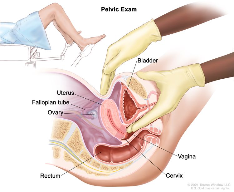 Pelvic exam. A doctor or nurse inserts one or two lubricated, gloved fingers of one hand into the vagina and presses on the lower abdomen with the other hand. This is done to feel the size, shape, and position of the uterus and ovaries. The vagina, cervix, fallopian tubes, and rectum are also checked.
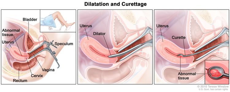 Dilatation and curettage (D and C). A speculum is inserted into the vagina to widen it in order to look at the cervix (first panel). A dilator is used to widen the cervix (middle panel). A curette is put through the cervix into the uterus to scrape out abnormal tissue (last panel).
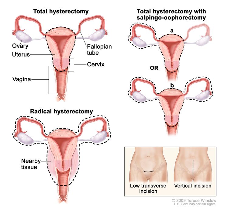 Hysterectomy. The uterus is surgically removed with or without other organs or tissues. In a total hysterectomy, the uterus and cervix are removed. In a total hysterectomy with salpingo-oophorectomy, (a) the uterus plus one (unilateral) ovary and fallopian tube are removed; or (b) the uterus plus both (bilateral) ovaries and fallopian tubes are removed. In a radical hysterectomy, the uterus, cervix, both ovaries, both fallopian tubes, and nearby tissue are removed. These procedures are done using a low transverse incision or a vertical incision.


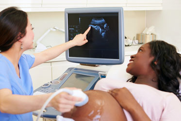 Ultrasound


 Medicurio