Osteo-articular tuberculosis

REQUEST AN APPOINTMENT OR BOOK A CONSULANT – Sargam.dange.18@gmail.com

Tuberculous synovitis frequently presents as a monoarthritis of weight-bearing joints such as the hip, knee, or ankle. Owing to its low incidence in developed countries, the diagnosis is often delayed for months to years. Early diagnosis with a synovial biopsy permits prompt antituberculous therapy and substantially improves the prospect of preservation of joint structure and function. Initial treatment typically includes combination therapy with four drugs (isoniazid, rifampin, pyrazinamide, and streptomycin or ethambutol) because of the frequency of isoniazid resistance. Antimicrobial therapy should be of at least 9 months’ duration, longer in immunocompromised hosts. Partial synovectomy and other surgical procedures should be restricted to joints with severe cartilage destruction, large abscesses, joint deformity, multiple drug resistance, or atypical mycobacteria.

Tubercular bacilli reach the joint space via the blood stream through subsynovial vessels, or indirectly from epiphyseal (more common in adults) or metaphyseal (more common in children) lesions, which erode into the joint space. Articular cartilage destruction begins peripherally and the weightbearing surfaces are preserved for a few months, providing the potential for good functional recovery with effective treatment in patients with early disease.

The disease may start in bone or in the synovial membrane, but one rapidly infects the other. The initial focus starts in the metaphysis in childhood or at the end of the bone in adults. An example of articular tuberculosis of the typical osseous areas of predilection for hip disease.

Osseous Changes and Tubercular Sequestra

After the infection, marked hyperemia and severe osteoporosis occur. The softened bone easily yields under the effect of gravity and muscle action, leading to compression, collapse, or deformation. Necrosis also may be caused by ischemic infarction of segments of bone.

Sequestration gives the appearance of coarse sand and rarely produces a radiologically visible sequestrum. Because of loss of nutrition, the adjacent articular cartilage or the intervening disc degenerate and also may become separated as sequestra. Some of the radiologically visible smaller sequestra in tuberculous cavities may result from calcification of caseous matter.

The Future Course of Tubercle

Before the availability of antitubercular drugs, the 5–year followup mortality of patients with osteoarticular tuberculosis was approximately 30.Modern antitubercular agents have changed the outlook considerably. Depending on the sensitivity pattern, host resistance, and the stage of the lesion at the inception of treatment, the tuberculous lesion may behave as follows. It may resolve completely; the disease may heal with residual deformity and loss of function; the lesion may be walled off completely and the caseous tissue may calcify; low-grade chronic fibromatous granulating and caseating lesion may persist (grumbling disease); and the infection may spread locally by contiguity and systematically by the blood stream

DIAGNOSIS OF TB INFECTION

Methods
Tuberculin Skin Test (TST)
Interferon Gamma Release Assays (IGRAs)
Enzyme-Linked Immunosorbent Assays (ELISA)
Bacteriology
Radiology
Computed Tomography (CT)
Magnetic Resonance Imaging (MRI)
Polymerase Chain Reaction (PCR)
Synovial fluid examination
Synovial biopsy

Blood

A relative lymphocytosis, low hemoglobin, and increased erythrocyte sedimentation rate often are found in patients with the active stage of disease. An increased erythrocyte sedimentation rate, however, is not necessarily proof of activity of the infection. Its repeated estimation at 3- to 6-month intervals gives an index of the activity of the disease.

Mantoux Test

As a rule, a positive reaction is present in a patient infected with tuberculosis for more than 1 month. A negative test, in general, rules out the disease. The tuberculin test rarely may be negative although active tuberculosis is present, such as in immune deficiency states.

Biopsy

Whenever there is doubt (particularly in the early stages) it is mandatory to prove the diagnosis by obtaining a biopsy specimen of the diseased tissue (granulations, synovium, bone, lymph nodes, or margins of tuberculous ulcers). Microscopic examination of an aspiration, core biopsy, needle biopsy, or open biopsy will reveal typical tubercles in patients who are not treated. Epithelioid cells surrounded by lymphocytes, even without central necrosis or peripheral foreign-body giant cells, are adequate histologic evidence of tuberculosis in a patient who is suspected to have the disease. At the time of open biopsy of a joint or bone, the orthopaedic surgeon should do therapeutic synovectomy or curettage. The infections of bone and joint that present as granulomatous lesions in order of frequency are tuberculosis, mycotic infection, brucellosis, sarcoidosis, and tuberculoid leprosy.

Guinea Pig Inoculation

The tuberculous pus, joint aspirate, or diseased material may be injected intraperitoneally into a guinea pig. Examination in positive cases shows tubercles on the peritoneum 5 to 8 weeks later. Although it currently is not considered a cost-effective test, it perhaps is the most reliable proof of tuberculosis.

Smear, Culture, and Serology

The material prepared for guinea pig inoculation also may be submitted for smear and culture examination for acid-fast bacilli. In superficial joints, one may be able to aspirate synovial fluid. Analysis of synovial fluid does not provide pathognomonic information; however, in general the leukocyte count is elevated to approximately 20,000 mm3, there is a lowered glucose level, and poor mucin. Clear synovial aspirate is not an appropriate material for microbiologic investigation; however, it is an excellent material for polymerase chain reaction and nucleic acid probes.

REQUEST AN APPOINTMENT OR BOOK A CONSULANT – Sargam.dange.18@gmail.com

Principles of Treatment of Osteoarticular Tuberculosis

Modern drugs promote the healing of sinuses, ulcers, and abscesses in patients previously unresponsive to extensive surgery. They also eliminate the danger of postoperative miliary and meningeal disease caused by dissemination of the tuberculous infection.

Death caused by uncontrolled disease, meningitis, miliary tuberculosis, amyloidosis, paralysis, and crippling now is rare. If a patient is diagnosed early and treated vigorously, healing can be accomplished without residual joint ankylosis or deformity.

With the use of modern drugs, the indications for surgery have become universally more selective and directed toward the prevention and correction of deformities, and the improvement in function of the diseased joints. At the stage of tuberculous arthritis, if abscess formation has not occurred, the natural outcome generally is a fibrous ankylosis. If an abscess discharges and sinuses develop, the outcome may be a bony ankylosis. The prognosis in articular tuberculosis depends on the stage of the disease. when the specific treatment is started. Concomitant disease must be treated and hospitalization is necessary only for patients with complications, or for patients requiring traction under supervision to correct deformities.

Category of treatmentCategory of TB casesAnti-TB drug regimens
Intensive phaseContinuation phase
INew Patient Regimen
New smear-positive PTB
Smear-negative PTB with extensive
Parenchymal involvement
Severe forms of EPTB other than TB meningitis
2HRZE4HR
IINew Patient Regimen
Smear-negative PTB without extensive
P Parenchymal involvement
Less severe forms of EPTB (e.g., TB cervical adenitis)
2HRZ4HR
IIINew Patient Regimen
TB meningitis
2HRZSa4HR
IVRetreatment regimen
Previously treated smear-positive PTB (relapse, treatment after interruption or treatment failure)
If low risk for MDR-TB or risk unknown, continue with retreatment regimen
If high risk for MDR-TB, use MDR-TB regimen below
2HRZES/1HRZE5HRE
VMDR Regimen
MDR-TB
Individualized regimens 

Table 2: Treatment regimens for tuberculosis recommended by the WHO . Other regimens are recommended for the treatment of TB meningitis, including replacing streptomycin with ethionamide and treating for 9-12 months.
E: Ethambutol; EPTB: Extra-Pulmonary Tuberculosis; H: Isoniazid; HIV: Human Immunodeficiency Virus; MDR-TB: Multi-Drug Resistant Tuberculosis; PTB: Pulmonary Tuberculosis; R: Rifampicin; S: Streptomycin; Z: Pyrazinamide; 2HRZ 4HR: Denotes a Two-month Intensive Phase of Daily Isoniazid, Rifampicin, and Pyrazinamide followed by a Four-month Continuation Phase of Daily Isoniazid and Rifampicin  

Rest, Immobilization, and Braces

In the active stage of disease, the joints are rested in the position of function using removable splints. Prolonged immobilization can lead to spontaneous ankylosis when joints are grossly destroyed. Patients with early disease are allowed 1 to 2 hours of intermittent, guarded active and assisted exercises while taking antitubercular drugs, with the aim of retaining a useful range of movement in the functional arc of the involved joint. Traction helps to correct deformity and to rest the diseased part. Gradual ambulation is encouraged with the help of suitable braces approximately 3 months after the start of treatment while healing is progressing. As the disease heals and pain subsides, weightbearing and activity are permitted. If there is steady progress, activity is increased within the limits of discomfort. The use of a brace is discontinued gradually after approximately 2 years.

Treatment of Abscess, Effusion, and Sinus

Palpable and large joint effusions are aspirated and 1000 mg of streptomycin alone or combined with injectable isoniazid (300 mg) is instilled at each aspiration. Local concentrations of antibiotics after parenteral administration may make this local instillation unnecessary. Open drainage of an abscess is indicated if aspiration fails. Radiologically visible paravertebral abscess shadows do not need to be drained, unless decompression is done in patients with paraplegia or when diseased vertebrae are debrided. A prevertebral abscess in the cervical region is drained if it causes difficulty in swallowing or breathing. Drainage of a large paravertebral abscess also may be considered when its radiologic size increases markedly despite treatment.

A majority of ulcers and sinuses heal within 6 to 12 weeks under the influence of systemic antitubercular drugs. Less than 1% of patients with sinuses require longer treatment and excision of the tract, with or without debridement. Sinus ramification always is greater than can be appreciated and complete surgical excision therefore is impractical.

REQUEST AN APPOINTMENT OR BOOK A CONSULANT – Sargam.dange.18@gmail.com

Antitubercular Chemotherapy

Combination chemotherapy should be used for an adequate length of time. Most of the antitubercular drugs potentially are toxic and resistance or intolerance to the drugs should be suspected when a patient fails to respond.

Multidrug Resistant Tuberculosis and Patients Who do not Respond

If the disease is caused by organisms resistant to isoniazid and rifampicin (multidrug resistant tuberculosis), if the disease is not controlled within 4 to 5 months, or if despite multidrug therapy, more active tuberculous lesions appear, one has to resort to second line drugs or potential antituberculous drugs. The situation is desperate in patients who do not respond. Immunomodulation in conjunction with drugs may be used in such patients. A favorable response was reported in approximately 85% of patients.

Pending the availability of better immunomodulation techniques.the current author has evolved the following outline during the past 12 years to upgrade cell-mediated immunity. In brief, 150 mg of levamisol is given at night for 3 days at weekly intervals for a total of 45 tablets. Four injections are administered once a month. The first and second infections are 0.1 mL intradermal (Bacillus Calmett-Guérin) injections and the third and fourth are intramuscular DPT injections (diphtheria vaccine + tetanus vaccine + Bordetella pertussis 20,000 million per 0.5 mL).

Surgery in Patients with Tuberculosis of Bones and Joints

No surgery is a substitute for a prolonged course of antitubercular drugs. A trial of conservative treatment is justified in most patients before surgery is contemplated. Nonoperative treatment usually is adequate in patients with pure synovial tuberculosis, low-grade or early arthritis of any joint, and even advanced (Stage III or IV) arthritis, especially in the upper extremity.

Surgery only should be considered once the general condition of the patient is stabilized by drug therapy, before the development of drug resistance. In general, a minimum of 1 to 4 weeks of therapy is advisable before any major surgical intervention.

Relapse of Osteoarticular Tuberculosis or Recurrence of Complications

The incidence of relapse or recurrence is unknown because these complications may occur at any period during the lifetime of a patient, whether the initial treatment included excisional surgery. Reactivation may occur in 2% to 5% of patients as late as 20 years or more after apparent healing.

The causes of reactivation include prolonged use of systemic cortisone therapy, malnutrition, the development of diabetes or an immune deficiency state, or a surgical procedure or injury to the previously infected area. Dormant bacilli persisting in tissue for years may start multiplying under such circumstances.

Tuberculosis of the Spine

Vertebral tuberculosis accounts for 50% of all cases of skeletal tuberculosis. In the majority of patients, the disease typically started in the paradiscal region. Narrowing of the disc often is the earliest radiologic finding. Any reduction in disc space, if it is associated with a loss of definition of the paradiscal margins of the vertebrae, suggests tuberculosis and occurs before the appearance of frank osseous destruction. These changes usually are evident only after infection has been present for 3 to 6 months, although advanced imaging may detect changes at approximately 6 weeks.

Most cases of tuberculosis of the spine heal without surgical intervention. However, uncertainty about the diagnosis, progressive bone destruction, and neurologic symptoms despite chemotherapy are definite indications for surgery in the active stage of disease.

REQUEST AN APPOINTMENT OR BOOK A CONSULANT – Sargam.dange.18@gmail.com

Tuberculosis: Spine( Pott’s Disease)

REQUEST AN APPOINTMENT OR BOOK A CONSULANT – Sargam.dange.18@gmail.com

Pott’s Disease, also known as tuberculosis spondylitis, is a rare infectious disease of the spine which is typically caused by an extraspinal infection. Pott’s Disease is a combination of osteomyelitis and arthritis which involves multiple vertebrae.  The typical site of involvement is the anterior aspect of the vertebral body adjacent to the subchondral plate and occurs most frequently in the lower thoracic vertebrae. A possible effect of this disease is vertebral collapse and when this occurs anteriorly, anterior wedging results, leading to kyphotic deformity of the spine.  Other possible effects can include compression fractures, spinal deformities and neurological insults, including paraplegia

Tuberculosis is an extremely infectious disease caused by the bacterium Mycobacterium tuberculosis. It’s one of the top-10 causes of death worldwide. Tuberculosis (TB) is most common in developing countries, but more than 9,000 cases were reported in the United States in 2016. Tuberculosis is preventable, and if it’s contracted and discovered early, it’s generally treatable.

TB primarily affects the lungs, but in some cases it can spread to other parts of the body. When TB spreads, it’s referred to as extrapulmonary tuberculosis (EPTB). One form of EPTB is bone and joint tuberculosis. Bone tuberculosis is simply a form of TB that affects the spine, the long bones, and the joints.

In the United States, only about 3 percent of all TB cases affect the musculoskeletal system. Of those cases, the spine is most commonly affected. Therefore, if you have bone TB, you are more likely to have it in or on your spinal column. However, bone TB could potentially affect any bone in your body. A common form of spinal bone TB is known as Pott’s disease.

Prevalence

Incidence
In 2005, there were 8.8 million new patients with tuberculosis (TB) all over the world, and of these, 7.4 million were in Asia and sub-Saharan Africa. Involvement of the spine reportedly occurs in less than 1-2% of patients who contract TB. Although the incidence of tuberculosis increased in the late 1980’s to early 1990’s, the total number of cases has decreased in recent years. In the United States, bone and soft tissue tuberculosis accounts for approximately 10% of extrapulmonary TB cases and between 1% and 2% of total cases. Of these cases, Pott’s disease is the most common manifestation of musculoskeletal TB, accounting for approximately 40-50%. Internationally, approximately 1-2% of total tuberculosis cases are attributable to Pott’s disease.

Ethnicity
Data from the United States show that musculoskeletal tuberculosis primarily affects African Americans, Hispanic Americans, Asian Americans, and foreign-born individuals.  The number of patients with TB spondylitis in Japan also declined to 233 in 2005 from 734 in 1978 and 276 in 2001.

Gender
Although some studies have found that Pott’s disease does not have sexual predilection, the disease is more common in males. The male to female ratio is reportedly 1.5-2:1.

Age
In the United States and other developed countries, Pott’s disease occurs primarily in adults. In underdeveloped countries which have higher rates of Pott’s disease, involvement in young adults and older children predominates.

causes

Tuberculosis is normally spread from person to person through the air. After you contract tuberculosis, it can travel through the blood from the lungs or lymph nodes into the bones, spine, or joints. Bone TB typically begins due to the rich vascular supply in the middle of the long bones and the vertebrae.

Bone tuberculosis is relatively rare, but in the last few decades the prevalence of this disease has increased in developing nations partially as a result of the spread of AIDS. While rare, bone tuberculosis is difficult to diagnose and can lead to severe problems if left untreated.

Characteristics/Clinical Presentation

Spinal Involvement

  • Lower thoracic vertebrae is the most common area of involvement (40-50%), followed by the Lumbar spine (35-45%)
  • Approximately 10% of Pott’s disease cases involve the cervical spine.
  • The thoracic spine is involved in about 65% of cases, and the lumbar, cervical and thoracolumbar spine in about 20%, 10% and 5%, respectively
  • The atlanto-axial region may also be involved in less than 1% of cases

Physical Findings

  • Localized Tenderness
  • Muscle Spasms
  • Restricted Spinal Motion
  • Spinal Deformity
  • Neurological Deficits

Back Pain

Back pain is the earliest and most common symptom. Patients with Pott’s disease usually experience back pain for weeks before seeking treatment and the pain caused by spinal TB can present as spinal or radicular. Although both the thoracic and lumbar spinal segments are nearly equally affected, the thoracic spine is frequently reported as the most common site of involvement. Together, thoracic and lumbar involvement comprise of 80-90% of spinal TB sites.

Neurological Signs

Neurologic abnormalities occur in 50% of cases and can include spinal cord compression with the following: 

  • Paraplegia
  • Paresis
  • Impaired sensation
  • Nerve root pain
  • Cauda equina syndrome

REQUEST AN APPOINTMENT OR BOOK A CONSULANT – Sargam.dange.18@gmail.com

Spinal Deformities

Almost all patients with Pott’s disease have some degree of spine deformity with thoracic kyphosis being the most common.

Constitutional Symptoms

  • Fever
  • Night sweats
  • Weight loss 
  • Malaise

Cervical Spinal TB

Cervical spine TB is a less common presentation occurring in approximately 10% of cases, but is potentially more serious because severe neurological complications are more likely. This condition is characterized by cervical pain and stiffness and symptoms can also include torticollis, hoarseness, and neurological deficits. Upper cervical spine involvement can cause rapidly progressive symptoms and neurologic manifestations occur early, ranging from a single nerve palsy to hemiparesis or quadriplegia. Retropharyngeal abscesses occur in almost all cases. In lower cervical spine insults, the patient can present with dysphagia or stridor.

Presentation in People Infected with HIV

The clinical presentation of spinal tuberculosis in patients infected with the human immunodeficiency virus (HIV) is similar to that of patients who are HIV negative; however, spinal TB seems to be more common in persons infected with HIV.

Asymptomatic Presentation

62-90% of patients with Pott’s disease are reported to have no evidence of extraspinal tuberculosis, further complicating a timely diagnosis.

Diagnostic Tests/Lab Tests/Lab Values

The Mantoux Test (Tuberculin Skin Test)
Injection of a purified protein derivative (PPD). Results are positive in 84-95% of patients with Pott’s disease who are not infected with HIV.

Erythrocyte Sedimentation Rate (ESR)
ESR may be markedly elevated (>100 mm/h)

Microbiology Studies
Microbiology studies are used to confirm diagnosis. Bone tissue or abscess samples are obtained to stain for acid-fast bacilli (AFB), and organisms are isolated for culture and susceptibility. CT-guided procedures can be used to guide percutaneous sampling of affected bone or soft tissue structures; however, these study findings are positive in only about 50% of the cases.

Radiography
Radiographic changes associated with Pott’s disease present relatively late. The following are radiographic changes characteristics of spinal tuberculosis on plain radiography:

  • Lytic destruction of anterior portion of vertebral body
  • Increased anterior wedging
  • Collapse of vertebral body
  • Reactive sclerosis on a progressive lytic process
  • Enlarged psoas shadow with or without calcification
  • Vertebral end plates may be osteoporotic
  • Intervertebral disks may be shrunk or destroyed
  • Vertebral bodies show variable degrees of destruction
  • Fusiform paravertebral shadows suggest abscess formation
  • Bone lesions may occur at more than one level

CT Scanning
CT scanning provides much better bony detail of irregular lytic lesions, sclerosis, disk collapse, and disruption of bone circumference. Low contrast resolution provides a better assessment of soft tissue, particularly in epidural and paraspinal areas. CT scanning reveals early lesions and is more effective for defining the shape and calcification of soft tissue abscesses which is common in TB lesions.

MRI
MRI is the criterion gold standard for evaluating disk-space infection and osteomyelitis of the spine and is most effective for demonstrating the extension of disease into soft tissue and the spread of tuberculous debris under the anterior and posterior longitudinal ligaments. MRI is also called the most effective imaging study for demonstrating neural compression. MRI findings useful to differentiate tuberculosis spondylitis from pyogenic spondylitis include thin and smooth enhancement of the abscess wall and well-defined paraspinal abnormal signal, whereas thick and irregular enhancement of abscess wall and ill-defined paraspinal abnormal signal suggest pyogenic spondylitis. Thus, contrast-enhanced MRI appears to be important in the differentiation of these two types of spondylitis.

Pott's Disease Abscess

               MRI of the thoracic spine (T2-weighted, sagittal reconstruction). The dorsal fluid collection suggests a 

          paravertebral abscess (large arrow) just above the fractured and operated third thoracic vertebra (small arrow)


Biopsy
Use of a percutaneous CT-guided needle biopsy of bone lesions can be used to obtain tissue samples. This is a safe procedure that also allows therapeutic drainage of large paraspinal abscesses.

Polymerase Chain Reaction (PCR)
PCR techniques amplify species-specific DNA sequences which is able to rapidly detect and diagnose several strains of mycobacterium without the need for prolonged culture. They have also been used to identify discrete genetic mutations in DNA sequences associated with drug resistance

Bone tuberculosis treatment

While bone tuberculosis can lead to some painful side effects, the damage is usually reversible when treated early with the right regimen of medications. In many cases, spinal surgery is necessary, such as a laminectomy (where a part of the vertebrae is removed).

Medications are the first line of defense for bone tuberculosis, and the course of treatment can last anywhere from 6–18 months. Treatments include:

  • antituberculosis medications, such as rifampicin, isoniazid, ethambutol and pyrazinamide
  • surgery

Medications

The duration of treatment is somewhat controversial. Although some studies favor 6 to 9 month course, traditional courses range from 9 months to longer than 1 year. The duration of therapy should be individualized and based on the resolution of active symptoms and the clinical stability of the patient.


The main drug class consists of agents that inhibit growth and proliferation of the causative bacteria. Isoniazid and rifampin should be administered during the whole course of therapy. Additional drugs are administered during the first two months of therapy and these are generally chosen among the first-line drugs which include pyrazinamide, ethambutol, and streptomycin. The use of second-line drugs is indicated in cases of drug resistance.


Isoniazid (Laniazid, Nydrazid)
Highly active against Mycobacterium tuberculosis. Has good GI absorption and penetrates well into all body fluids and cavities.


Rifampin (Rifadin, Rimactane)
For use in combination with at least one other antituberculous drug; inhibits DNA-dependent bacterial but not mammalian RNA polymerase. Cross-resistance may occur.


Pyrazinamide
Bactericidal against M tuberculosis in an acid environment (macrophages). Has good absorption from the GI tract and penetrates well into most tissues, including CSF.


Ethambutol (Myambutol)
Has bacteriostatic activity against M tuberculosis. Has good GI absorption. CSF concentrations remain low, even in the presence of meningeal inflammation.


Streptomycin
Bactericidal in an alkaline environment. Because it is not absorbed from the GI tract, must be administered parenterally. Exerts action mainly on extracellular tubercle bacilli. Only about 10% of the drug penetrates cells that harbor organisms. Enters the CSF only in the presence of meningeal inflammation. Excretion is almost entirely renal.

Physical Therapy Management (current best evidence)

Patients with Pott’s disease often undergo spinal fusion or spinal decompression surgeries to correct their structural deformity and prevent further neurological complications. There are no established guidelines which dictate treatments that will yield positive outcomes in such patients.  However, treatment regimens should address each patient individually, focusing on any impairments, functional limitations and/or disabilities with which they present.

PT Managment Post-Spinal Decompression Surgery

  • Spinal Stabilization Exercises
  • Maitland
  • Back School
  • Exercise and Strengthening

When compared with other physical therapy treatments and self-managment, spinal stabilization exercises were found to produce significantly more positive ratings in global outcomes. Pain and disability, however, did not show significant improvement when compared to the other two treatment options.

PT Managment Post-Spinal Fusion Surgery

  • TENS (Transcutaneous Electrical Neuromuscular Stimulation)
  • Aquatic Therapy
  • Overground Training (Walking Program)
  • Aerobic Exercise
  • Trunk Strengthening

Studies examining the use of TENS have shown higher frequencies are more effective in decreasing neuropathic pain. Aerobic exercise, PT, and trunk strengthening interventions have all attained significant decreases in pain, psychological distress and disability.

REQUEST AN APPOINTMENT OR BOOK A CONSULANT – Sargam.dange.18@gmail.com

Volkamann’s Ischaemic Contracture

REQUEST AN APPOINTMENT OR BOOK A CONSULANT – Sargam.dange.18@gmail.com

Volkmann contracture is a deformity of the hand, fingers, and wrist caused by injury to the muscles of the forearm. The condition is also called Volkmann ischemic contracture.

A Volkmann’s contracture is deformity of the hand, fingers, and wrist which occurs as a result of a trauma such as fractures, crush injuries, burns and arterial injuries. Following this trauma, there is a deficit in the arterio-venous circulation in the forearm which causes a decreased blood flow and hypoxia can lead to the damage of muscles, nerves and vascular endothelium. This results in a shortening (contracture) of the muscles in the forearm.

Causes

Volkmann contracture occurs when there is a lack of blood flow (ischemia) to the forearm. This occurs when there is increased pressure due to swelling, a condition called compartment syndrome.

Injury to the arm, including a crush injury or fracture, can lead to swelling that presses on blood vessels and decreases blood flow to the arm. A prolonged decrease in blood flow injures the nerves and muscles, causing them to become stiff (scarred) and shortened.

When the muscle shortens, it pulls on the joint at the end of the muscle just as it would if it were normally contracted. But because it is stiff, the joint remains bent and stuck. This condition is called a contracture.

In Volkmann contracture, the muscles of the forearm are severely injured. This leads to contracture deformities of the fingers, hand, and wrist.

There are three levels of severity in Volkmann contracture:

  • Mild — contracture of 2 or 3 fingers only, with no or limited loss of feeling
  • Moderate — all fingers are bent (flexed) and the thumb is stuck in the palm; the wrist may be bent stuck, and there is usually loss of some feeling in the hand
  • Severe — all muscles in the forearm that both flex and extend the wrist and fingers are involved; this is a severely disabling condition. There is minimal movement of the fingers and wrist.

Conditions that can cause increased pressure in the forearm include:

  • Animal bites
  • A forearm fracture
  • Bleeding disorders
  • Burns
  • Injection of certain medicines into the forearm
  • Injury of the blood vessels in the forearm
  • Surgery on the forearm
  • Excessive exercise — this would not cause severe contractures

Signs and symptoms

The clinical presentation includes the five Ps:

  • Pain (earliest manifestation), especially accentuated by passive stretching
  • Pallor
  • Pulselessness
  • Paresthesias
  • Paralysis

Additional useful findings are as follows:

  • Firmness of the tissues on palpation
  • Induration of the forearm

The clinical presentation of Volkmann`s contracture includes what is commonly referred to as the 5 Ps. These are pain, pallor, pulselessness, paresthesias, and paralysis. Pain is the earliest sign

Special findings:

  • Bleach view at the level of the skin (pallor).
  • The wrist is in palmar flexion
  • Clawed fingers
  • Pain occurs with passive stretching of the flexor
  • Palpation of the affected region creates persistent pain (pain)
  • It is possible that the pulsations can not be felt in the swollen arm, mainly in the distal part (pulselessness).
  • There are also neurological limitations noticeable from the muscles that pinch the neural pathways, there is a decreased sensation (paresthesia) and there is an observable motor deficit (paresis)

REQUEST AN APPOINTMENT OR BOOK A CONSULANT – Sargam.dange.18@gmail.com

Diagnostic Procedures

Pressure monitoring

Intracompartmental pressure (ICP) can be measured by several means including:

  • Wick catheter
  • Simple needle manometry
  • Infusion techniques
  • Pressure transducers
  • Side-ported needles

Critical pressure for diagnosing compartment syndrome is unclear

Different authors consider surgical intervention if:

  • Absolute ICP greater than 30 mmHg
  • Difference between diastolic pressure and ICP greater than 30 mmHg
  • Difference between mean arterial pressure and ICP greater than 40 mmHg

Differential Diagnosis

Pseudo-Volkmann’s contracture

Examination

For a Volkmann’s contracture, the findings are specific as described in the clinical presentation subheading above. The main physical picture that we see is a neurological deficit that occurs in the nerves that pass in the affected regions. The flexion of the wrist is a result of contracture and a loss of innervation.

The deformity seen in this condition can be divided into different levels of severity:

  1. MILD: Flexion contracture of 2 or 3 fingers with no or limited loss of sensation
  2. MODERATE: All fingers are flexed and the thumb is oriented in the palmar orientation. The fist, in this case, can remain permanently flexed and there is usually a loss of sensation in the hand.
  3. SERIOUS: All muscles in the forearm (flexors and extensors) are involved. This is a serious limiting condition.

An objective test to evaluate the ischemia and the pressure in a muscle compartment is an invasive test. It measures the absolute pressure in the compartment of the muscle. This is also called the intracompartimental pressure monitoring (ICP)

Medical Management

Prevention is the best management in this condition. However, there are times, that surgical intervention will be indicated. The majority of Volkmann’s contractures are caused by a supracondylar fracture, and it is essential that all steps are taken to improve the healing of the fracture. When there is an intra-compartment pressure (ICP) of >30 mmHg,an urgent fasciotomy is recommended to avoid further complications, Raised ICP threatens the viability of the limb and compartment syndrome (CS) represents a true medical emergency. Thus, the need for decompression by removal of all dressing down to the skin, followed by fasciotomy- Surgical opening of the fascia around the muscles to make more place for the structures inside. This is done to prevent the onset of Volkmann’s contractures.

In moderate Volkmann’s contracture, tendon slide and neurolysis surgery should be performed (median and ulnar) along with extensor transfer procedures.

Finally, in severe cases of Volkmann’s contracture, debridement of injured muscle may be performed with releases of scar tissue and salvaging procedures. Range of motion and function after injury are improved by physical and occupational therapy.

Physiotherapy Management

Dynamc splint.jpg

After the surgery, it is important to ensure that the mobility is recovered by:

  • Passive stretching techniques
  • Range of motion exercises to enhance soft tissue elasticity.

Another part of the therapy programme involves activating and strengthening the weak agonist to ensure equilibrium in agonist and antagonist pull during joint movement.

Progressive Splinting, passive stretching and tendon gliding, as well as massage can be used in mild to moderate cases of Volkmann’s contracture.

By the use of an electromyographic device, the patient can train its affected muscles with cooperativity. The patient is more alert and there is more interaction between the patient and the therapist.

REQUEST AN APPOINTMENT OR BOOK A CONSULANT – Sargam.dange.18@gmail.com

Monteggia fracture dislocation

REQUEST AN APPOINTMENT OR BOOK A CONSULANT – Sargam.dange.18@gmail.com

Monteggia fracture-dislocations consist of a fracture of the ulnar shaft with concomitant dislocation of the radial head. The ulnar fracture is usually obvious, whereas the radial head dislocation can be overlooked, with potentially serious functional and medico-legal ramifications. 

The eponym Monteggia fracture is most precisely used to refer to a dislocation of the proximal radioulnar joint (PRUJ) in association with a forearm fracture, most commonly a fracture of the ulna. These injuries are relatively uncommon, accounting for fewer than 5% of all forearm fractures. The mechanism of injury is most often a fall on an outstretched hand.

The ulna fracture is usually clinically and radiographically apparent. Findings associated with the concomitant radial head dislocation are often subtle and can be overlooked. The keys to successful diagnosis of a Monteggia fracture are clinical suspicion and radiographs of the entire forearm and elbow. Properly assessing the nature of this injury in a timely fashion is imperative in order to prevent permanent disability or limb dysfunction.

Anatomy

The annular and radial collateral ligaments stabilize the radial head. These ligaments stretch or rupture during radial head dislocation. The radial head articulates with the humeral capitellum and the radial notch of the proximal ulna. The radius and ulna are closely invested by the interosseous membrane, which accounts for the increased risk of displacement or injury to the radius when the ulna fractures.

The distal ulna and radius also articulate at the DRUJ. The ulna provides a stable platform for rotation of the radius and forearm. The ulna and interosseous membrane also may provide stable platforms for dislocation of the proximal radius, leading to the Monteggia fracture.

The posterior interosseous nerve travels around the neck of the radius and dives under the supinator as it courses into the forearm. The median and ulnar nerves enter the antecubital fossa just distal to the elbow. The close proximity of these nerves may lead to injuries when a Monteggia fracture occurs. Neural injuries are generally traction injuries and result from stretching around the displaced bone or from energy dispersed during the initial injury.

Etiology

Monteggia fractures are primarily associated with falls on an outstretched hand with forced pronation. If the elbow is flexed, the chance of a type II or III lesion is greater. In some cases, a direct blow to the forearm can produce similar injuries.

Evans in 1949 and Penrose in 1951 studied the etiology of Monteggia fractures on cadavers by stabilizing the humerus in a vise and subjecting different forces to the forearm. Penrose considered type II lesions a variation of posterior elbow dislocation. Bado believed that the type III lesion, the result of a direct lateral force on the elbow, was primarily observed in children.

In essence, high-energy trauma (eg, a motor vehicle collision) and low-energy trauma (eg, a fall from a standing position) can result in the described injuries. A high index of suspicion, therefore, should be maintained with any ulna fracture.

Pathophysiology

The forearm structures are intricately related, and any disruption to one of the bones affects the other. The ulna and radius are in direct contact with each other only at the PRUJ and the DRUJ; however, they are unified along their entire length by the interosseous membrane. This allows the radius to rotate around the ulna. When the ulna is fractured, energy is transmitted along the interosseous membrane, displacing the proximal radius. The end result is a disrupted interosseous membrane proximal to the fracture, a dislocated PRUJ, and a dislocated radiocapitellar joint.

Radial head dislocation may lead to radial nerve injury. The posterior interosseous branch of the radial nerve, which courses around the neck of the radius, is especially at risk, particularly in Bado type II injuries. Injuries to the anterior interosseous branch of the median nerve and the ulnar nerve also have been reported. Most nerve injuries are neurapraxias and typically resolve over a period of 4-6 months. Splinting of the wrist in extension and finger range-of-motion (ROM) exercises help prevent contractures from developing while the patient awaits resolution of the nerve injury.

Classification

In 1814, Giovanni Battista Monteggia of Milan first described this injury as a fracture to the proximal third of the ulna with associated anterior dislocation of the radial head. Interestingly, he described this injury pattern in the pre-Roentgen era solely on the basis of the history of injury and the physical examination findings. However, this particular fracture pattern only accounts for about 60% of these types of injuries.

More than 150 years later, in 1967, Bado coined the term Monteggia lesion and classified the injury into the following four types:

  • Type I – Fracture of the proximal or middle third of the ulna with anterior dislocation of the radial head
  • Type II – Fracture of the proximal or middle third of the ulna with posterior dislocation of the radial head
  • Type III – Fracture of the ulnar metaphysis with lateral dislocation of the radial head
  • Type IV – Fracture of the proximal or middle third of the ulna and radius with anterior dislocation of the radial head

REQUEST AN APPOINTMENT OR BOOK A CONSULANT – Sargam.dange.18@gmail.com

All four types of Monteggia fracture-dislocations (see Bado classification) are treated with open reduction and internal fixation of the ulna and radius in type 4. Usually anatomical reduction of the ulna allows for a closed reduction of the radial head.  Types I, III, IV are cast at 110 degrees of flexion, whereas type II is cast at 70 degrees of flexion 4,6.

In the pediatric population, treatment depends on the type of ulnar fracture: 7,8

  • bowing/greenstick fracture: closed reduction of the ulnar bow and casting. If reduction of the radial head is not possible open osteotomy and fixation of the ulna may be required
  • complete transverse/short oblique fracture: closed reduction and intramedullary fixation
  • long oblique/comminuted fracture: open reduction and internal fixation

The prognosis is significantly decreased by delayed diagnosis and treatment.

Approach Considerations

Pain should be managed as needed in the immediate period. If the fracture is open, the status of the patient’s tetanus immunization should be determined and addressed as indicated. Intravenous (IV) antibiotics should be administered to patients with open fractures. Open wounds should be irrigated with sterile saline solution and dressed with sterile, moist gauze. The radial head should be reduced in the emergency department (ED) if possible.

Nonoperative treatment is successful for most Monteggia injuries in children, for the following reasons :

  • The majority of the fractures are inherently stable
  • Children require a shorter time for both the osseous and the ligamentous injuries to heal
  • Children have little trouble regaining motion lost through stiffness, despite immobilization of the fractures for the duration of the initial healing period (3-6 weeks)
  • The potential may exist for remodeling of mild residual angular deformities (< 10°)

Indications for treatment of Monteggia fractures are based on the specific fracture pattern and the age of the patient (ie, pediatric or adult).  Although most pediatric fracture patterns can be managed conservatively with closed reduction and long arm casting, most adult fractures require open reduction and internal fixation (ORIF).  Few contraindications for surgery exist. Once the radial head is reduced in closed injuries, surgical treatment may be delayed until the patient is stable and the surgery may be performed in a more elective fashion.

Surgical Therapy

Open fractures require emergency surgical consultation. The initial treating physician may reduce the radial head dislocation and splint this fracture. Otherwise, an orthopedic surgeon should be consulted immediately to reduce the radial head. Anatomic reduction of the ulna is usually required before radial head reduction. Unless the fracture is open, surgical treatment is performed on an elective basis. Whereas most adults require operative treatment, most pediatric fractures are treated with closed reduction.

Operative fixation of complete fractures of the ulna with proximal radioulnar joint (PRUJ) dislocation is recommended in children. The complete disruption of bone continuity is likely to be associated with substantial soft-tissue trauma in these injuries. Shortening and angulation of complete fractures after cast immobilization is not uncommon. Anatomic reduction of the ulnar fracture and radial head often requires operative treatment.

REQUEST AN APPOINTMENT OR BOOK A CONSULANT – Sargam.dange.18@gmail.com

Supracondylar Humeral Fracture physiotherapy approach

REQUEST AN APPOINTMENT OR BOOK A CONSULANT – Sargam.dange.18@gmail.com

Diagnostic Procedures

Displaced Supracondylar fracture in X-ray

Radiographs should include true AP of distal humerus (not elbow) and true lateral elbow views.If there is only minimal or no displacement these fractures can be occult on radiographs. The only sign will be a positive fat pad sign.

On lateral view

The lateral view also allows assessing the degree of displacement and the integrity of the posterior cortex.

On the lateral view, the following radiological parameters are looked for: (a) Anterior humeral line; (b) Coronoid line; (c) Fish tail sign; (d) Fat pad sign; (Anterior and Posterior).

Anterior Humeral Line Abnormal

Anterior humeral line to capitulum orientation on a lateral view

  • Normal elbow – line continues the anterior cortical of the humerus and should traverse the capitulum in its middle third line.
  • Extension type injury: capitulum posterior to line
  • Flexion type injury: capitulum anterior to the line

On AP view

It helps to evaluate the direction of displacement, the presence of varus or valgus alignment, and the extent of the fracture comminution.

The Baumann angle (Humeral capitular angle) on AP view

  • Angle between linea perpendicular to longthe axis of humeral shaft and the physeal line of the capitulum, isused to assess varus or valgus alignment of the distal humerus.
  • Normal range – 64º to 82º degrees
  • Decrease in angle – varus angulated fracture with possible medial column comminution

The Ulnohumeral angle or radiological carrying angle on AP view

  • The angle formed by the diaphyseal axis of the humerus and the axis of the proximal third of the ulna.
  • It is also used to assess varus or valgus deformity and it is more accurate and useful than Baumann’s angle 

Management / Interventions

Medical

Management of supracondylar fracture is determined based on the type of fracture based on Modified Gartland Classification.


Type I ( Non-displaced fracture)

  • Immobilization with a long-arm cast or splint.
  • With elbow flexion up to 80° to 90° and mid pronation-supination are well toleratable for ~3weeks.
  • Flexion of the elbow within the cast should not pass 90° because it can increase forearm pressures and impede distal vascular flow.
  • Radiographic check at 1 and 2 weeks

Types II

  • A closed reduction(CR) and percutaneous pinning fixation is recommended than CR and immobilization as the risk of complication are low. Pins are removed in the hospital approximately three weeks after surgery.

Type III and Type IV

  • Closed reduction and percutaneous pinning is the gold standard for all displaced fractures and is widely used in Type III and IV fractures.
  • Open reduction is indicated:
  1. When the surgeon is not able to reduce the fracture by closed means
  2. When there is soft-tissue entrapment (i.e. muscle, median nerve, brachial artery) or
  3. When a cold hand remains without perfusion after an attempt at closed reduction has been performed.

There is an increased incidence of infection, stiffness, and myositis ossificans in open reduction. The anterior approach is the most widely used approach for open reduction mainly when vascular repair is necessary. The lateral approach is standard for elbow surgery but in supracondylar fracture increases the risk of radial nerve injury and stiffness. The bilaterotricipital posterior approach (Alonso-Llames approach) is not recommended as it has a high rate of complications described, such as stiffness, unsightly scarring and risk of trochlea osteonecrosis.

REQUEST AN APPOINTMENT OR BOOK A CONSULANT – Sargam.dange.18@gmail.com

Physiotherapy Management

Physiotherapy treatment is vital in all patients with a supracondylar fracture to hasten healing and ensure an optimal outcome. The goal of physiotherapy treatment is:

  • To achieve painless and full mobility of the elbow joint.
  • To enhance healing process.
  • To strength the affected musculature.
  • To improve overall functional abilities of children.

Outcome measures that can be used to compare and evaluate the outcome of treatment are :

  • Numerical pain rating scale (NPRS)/ Faces Pain Scale
  • Range of motion- Goniometry
  • Manual muscle testing ( MMT)
  • ASK-p (Activities Scale for Kids-performance version)
  • Flynn’s criteria include two factors ‘cosmetic factor’ (loss of carrying angle) and ‘Functional factor’ (motion loss in degrees).
  • Neuro-vascular assessment is must post-operatively and during rehabilitation.

Evidence

The physical therapy management in the pediatric population is very controversial, in both its effect and its necessity. A randomized controlled trial was done by Schmale et al. in 2014, had shown that children with supracondylar fracture treated with either casting or CR with percutaneous pining followed by casting were not benefited by a short course of physical therapy (six sessions of physical therapy performed over a five-week period beginning the week after cast removal) in terms of either return of function or motion.Supporting the above study, another study was done in 2018, also shows that children managed with CR for uncomplicated supracondylar fracture with immobilization for three weeks regain their functional ROM within 12 weeks of mobilization by themselves, with no added benefit from physiotherapy.

Physiotherapy treatment has not shown significant difference, it may be due to the involvement of children of a ( 5- 10 years) in more involved in daily household activities from an early period and urge for a motion for playing. It would be either affected as the therapist may have been unduly aggressive or unduly conservative while providing treatment.

On the contrary, in a more severe type of injury with neurovascular involvement and in an adult patient, physiotherapy treatment has a significant role.And in the pediatric population, evidence has been lacking to address the strength of upper extremity as an outcome after physiotherapy treatment.

Therefore,

  • Optimal loading(pain-free activities based on a child) is very necessary for the pediatric population with supracondylar fracture as pain aggravating activities may delay the healing process and cause further damage as they are in the growing phase.
  • Thus, active exercise and active involvement in sports and ADLs are recommended rather than passive joint mobilization and stretching exercises.
  • Those activities such as lifting, weight-bearing, or pushing activities that pose large amounts of stress through the humerus should also be avoided in the initial week after immobilization removable.
  • Progressive strengthening exercises can be addressed.

Advice and Exercises During the Immobilization Period

  • Normally elbow is immobilized for 3 weeks so, during that period, adjacent joints ( Shoulder joint and wrist joint and hand) should be kept moving active or active-assisted exercises frequently in a day.
  • Elbow shouldn’t be moved and used of arm sling should be proper.
  • Postural training( sitting upright with relaxed shoulder and retraction of the scapula) should be taught.

1-2 Weeks After Removal of Cast

  • Hot fermentation can be used to ease joint stiffness.
  • Gentle soft tissue release can be done in arm and forearm musculature.
  • Gentle active and active-assisted exercises using a wand and be taught in pain- free limit- frequently in a day.
  • Isometric exercises for arm and forearm musculature can be addressed.
  • Educate parents and child to use the affected hand to use in daily activities like brushing, writing, eating, dressing, etc.
  • Avoid weight lifting and pushing activities.
12973256(300x300).jpg
12974256(300x300).jpg

Advice and Exercise After 2 Weeks of Cast Removal

  • Progressive and interactive range of motion exercise and strengthening exercise should be addressed eg: passing a ball, dressing and undressing clothes.

Conclusion

  • Supracondylar fractures of the humerus are the most frequent fractures in children with a peak incidence at the ages of five to eight years.
  • FOOSH is the most mechanism of injury of Supracondylar fracture of the humerus.
  • The Neurovascular assessment is must pre and post-operatively.
  • Closed reduction with percutaneous pinning is the recommended medical management for displaced fracture without neurovascular involvement.
  • Active exercise is recommended in pediatric elbow fracture rather than passive treatment.

REQUEST AN APPOINTMENT OR BOOK A CONSULANT – Sargam.dange.18@gmail.com

fracture shaft humerus

REQUEST AN APPOINTMENT OR BOOK A CONSULANT – Sargam.dange.18@gmail.com

Humeral shaft fractures are common injuries. Like many orthopaedic injuries, they have a bimodal distribution, occurring in both younger patients due to high energy trauma and in elderly patients following low impact injuries.

Due to the location of the radial nerve within the spiral groove, there is a reasonably high risk of injury; the overall incidence is around 10%, although this is much higher (~25%) in Holstein-Lewis fractures (as discussed below).

The risk factors for humeral shaft fractures include osteoporosis, increasing age, or previous fractures. Humeral shaft fractures can also occur as pathological fractures.

Anatomy
Osteology humeral shaft is cylindrical distally humerus becomes triangular intramedullary canal terminates 2 to 3 cm proximal to the olecranon fossa
Muscles insertion for pectoralis major  deltoid  coracobrachialis  origin for  brachialis  triceps  brachioradialis 
Nerve radial nerve  courses along spiral groove  14cm proximal to the lateral epicondyle 20cm proximal to the medial epicondyle

Humeral shaft fractures account for 3-5% of all fractures . Although they occur in all age groups, a bimodal distribution is noted. The first peak is seen in the third decade in males and the second peak in the seventh decade in females .

causes

A broken arm is a common injury and is usually a consequence of a fall with an outstretched hand, a car crash or some other type of accident.

Clinical Features

Pain and deformity are the predominant features of this injury. These fractures may occur from a fall directly onto the outstretched limb or falling laterally onto an adducted limb.

If the radial nerve is involved, the patient may also complain of reduced sensation over the dorsal 1st webspace and weakness in wrist extension.

On examination, ensure you carefully check and document the neurovascular status*. Assess for open wounds and any suspected concurrent injuries or fractures, particularly if there was a high-energy impact involved.

Classification
OTA bone number
: 1 fracture location:
2 fracture pattern:
simple:A,
wedge:B,
complex:C
Descriptive
fracture location: proximal,
middle or distal third
fracture pattern: spiral,
transverse,
comminuted
Holstein-Lewis fracture   a spiral fracture of the distal one-third of the humeral shaft commonly associated with neuropraxia of the radial nerve (22% incidence)

Holstein-Lewis Fracture

A Holstein-Lewis fracture is a fracture of the distal third of the humerus resulting in the entrapment of the radial nerve.

The resultant neuropraxia to the radial nerve will result in loss of sensation in the radial distribution and a wrist drop deformity. Surgical management is indicated in such cases.

symptoms

Symptoms vary depending on the specific type of fracture but may include:

  • Pain
  • Swelling and bruising
  • Inability to move the shoulder
  • A grinding sensation when the shoulder is moved
  • Deformity — “It does not look right.”
  • Occasionally bleeding (open fracture)
  • Loss of normal use of the arm if a nerve injury occurs 

REQUEST AN APPOINTMENT OR BOOK A CONSULANT – Sargam.dange.18@gmail.com

Investigations

Anteroposterior (AP) and lateral plain film radiographs of the humerus are usually that is all that is required (Fig. 2). The elbow and shoulder should be visible.

In severely comminuted cases, CT imaging may be requested for pre-operatively planning, although this is not routinely done.

Complications

In most cases the prognosis is good, with minimal impact on function once the fracture has healed. Non-union and mal-union are important, albeit fortunately rare, complications to consider in humeral shaft fractures.

Varus angulation is slightly more common with transverse fractures, however rarely causes functional limitations, as the shoulder has such a vast range of motion that most deformities in the humerus can be accommodated for.

Around 90% of radial nerve injuries will improve within 3 months without any intervention.

treatment for a humerus fracture

Proximal Humeral Fracture

Most fractures of the proximal humerus can be treated without surgery if the bone fragments are not shifted out of position (displaced). If the fragments are shifted out of position, surgery is often performed to allow earlier mobility. However, other factors are also considered when deciding between surgical fixation or nonoperative treatment. 

Nonoperative treatment is usually with a sling or shoulder immobilizer with no shoulder mobility for the first two weeks. Thereafter, the patient will be given weekly exercises to slowly increase the shoulder’s range of motion. An X-ray of the shoulder will be taken on a weekly or biweekly (every two weeks) basis to confirm the fracture is healing properly. 

Surgery usually involves fixation of the fracture fragments with plates, screws or pins. Severe fractures with previous arthroscopy (joint degeneration) may require shoulder replacement. Mobilization with physical therapy is begun immediately following surgery. 

Humerus Shaft Fracture

A humerus shaft fracture may be treated with or without surgery, depending on the fracture pattern and associated injuries (i.e., nerve injury or open fracture). A temporary splint extending from the shoulder to the forearm and holding the elbow bent at 90 degrees can be used for initial management of the fracture.

Nonoperative treatment usually includes the placement of fracture bracing that will be replaced by a cylindrical brace (Sarmiento brace) three to four weeks later that fits the upper arm while leaving the elbow free. The doctor will tell you how long to wear the cast or splint and will remove it at the right time. It may take several weeks to several months for the broken arm to heal completely. 

Rehabilitation involves gradually increasing activities to restore muscle strength, joint motion and flexibility. The patient’s cooperation is essential to the rehabilitation process. The patient must complete range of motion, strengthening and other exercises prescribed by the doctor on a daily basis. Rehabilitation will continue until the muscles, ligaments and other soft tissues perform normally. 

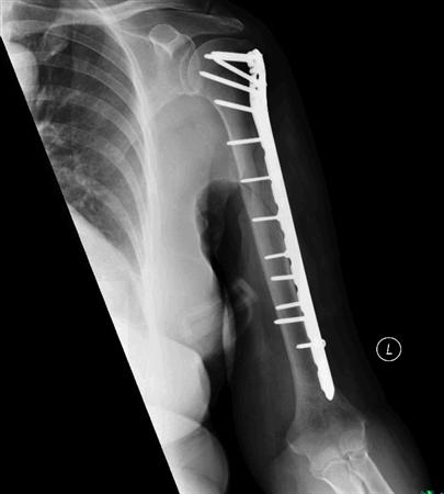
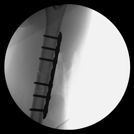
Surgery usually involves internal fixation of the fragments with plates, screws or a nail. The rehabilitation differs slightly from nonoperative treatment, with no splints or cast. The patient is usually given a sling for comfort and arm support. Elbow exercises may be started immediately after surgery, while shoulder exercises may be delayed for a few weeks based on the fracture pattern.

Nonoperative
  coaptation splint followed by functional brace 
indications
indicated in vast majority of humeral shaft fractures criteria for acceptable alignment include: 
< 20° anterior angulation
< 30° varus/valgus angulation
< 3 cm shortening absolute
contraindications severe soft tissue injury or bone loss vascular injury requiring repair brachial plexus injury
relative contraindications  relative operative indications section radial nerve palsy is NOT a contraindication to functional bracing 
outcomes
90% union rate
  increased risk with proximal third oblique or spiral fracture  varus angulation is common but rarely has functional or cosmetic sequelae
damage control orthopaedics (DCO)
closed humerus fractures, including low velocity GSW, should be initially managed with a splint or sling  
type of fixation after trauma should be directed by acceptable fracture alignment parameters, fracture pattern and associated injuries
Operative
  open reduction and internal fixation (ORIF) 
absolute indications 
open fracture 
vascular injury requiring repair
brachial plexus injury 
ipsilateral forearm fracture (floating elbow)  
compartment syndrome
periprosthetic humeral shaft fractures at the tip of the stem
relative indications bilateral humerus fracture
polytrauma or associated lower extremity fracture 
allows early weight bearing through humerus
pathologic fractures burns or soft tissue injury that precludes bracing fracture characteristics
distraction at fracture site 
short oblique or transverse fracture pattern
intraarticular extension
intramedullary nailing (IMN) 
relative indications
pathologic fractures
segmental fractures
severe osteoporotic
bone overlying skin
compromise limits open approach 
polytrauma
Techniques
Coaptation Splint & Functional Bracing
coaptation splint 
applied until swelling resolves adequately
applied splint will extend up to axilla and over shoulder
common deformities include varus and extension valgus mold to counter varus displacement functional bracing  extends from 2.5 cm distal to axilla to 2.5 cm proximal to humeral condyles sling should not be used to allow for gravity-assisted fracture reduction
shoulder extension used for more proximal fractures weekly radiographs for first 3 weeks to ensure maintenance of reduction every 3-4 weeks after that
Open Reduction and Internal Fixation (ORIF)
approaches
anterolateral approach to humerus  used for proximal third to middle third shaft fractures  distal extension of the deltopectoral approach radial nerve identified between the brachialis and brachioradialis distally posterior approach to humerus   used for distal to middle third shaft fractures although can be extensile
triceps may either be split or elevated with a lateral paratricipital exposure

radial nerve is found medial to the long and lateral heads and 2cm proximal to the deep head of the triceps 
radial nerve exits the posterior compartment through lateral intramuscular septum 10 cm proximal to radiocapitellar joint 
lateral brachial cutaneous/posterior antebrachial cutaneous nerve serves as an anatomic landmark leading to the radial nerve during a paratricipital approach
 
techniques
plate osteosynthesis commonly with 4.5mm plate (narrow or broad)
3.5mm plates may function adequately
absolute stability with lag screw or compression plating in simple patterns 
apply plate in bridging mode in the presence of significant comminution  postoperative
full crutch weigh
t bearing shown to have no effect on union 
Intramedullary Nailing (IMN)
techniques
can be done antegrade or retrograde complication  
nonunion
nonunion rates not shown to be different between IMN and plating in recent meta-analyses 
IM nailing associated with higher total complication rates   
shoulder pain
increased rate when compared to plating (16-37%) 
functional shoulder outcome scores (ASES scores) not shown to be different between IMN and ORIF 
nerve injury  while controversial, a recent meta-analysis showed no difference between the incidence of radial nerve palsy between IMN and plating 
radial nerve is at risk with a lateral to medial distal locking screw 
musculocutaneous nerve is at risk with an anterior-posterior locking screw  postoperative
full weight bearing allowed and had no effect on union

REQUEST AN APPOINTMENT OR BOOK A CONSULANT – Sargam.dange.18@gmail.com

Management of fracture calvicle (Broken Collarbone)

REQUEST AN APPOINTMENT OR BOOK A CONSULANT – Sargam.dange.18@gmail.com

A clavicle fracture is a break in the collarbone, one of the main bones in the shoulder. This type of fracture is fairly common—accounting for about 5 percent of all adult fractures. Most clavicle fractures occur when a fall onto the shoulder or an outstretched arm puts enough pressure on the bone that it snaps or breaks. A broken collarbone can be very painful and can make it hard to move your arm.

Most clavicle fractures can be treated by wearing a sling to keep the arm and shoulder from moving while the bone heals. With some clavicle fractures, however, the pieces of bone move far out of place when the injury occurs. For these more complicated fractures, surgery may be needed to realign the collarbone.

The clavicle is located subcutaneously between the sternum and the scapula, and it connects the arm to the body.

ClavicleFracture2.jpg

 The clavicle is the first bone in the human body to begin intramembranous ossification directly from mesenchyme during the fifth week of fetal life. Similar to all long bones, the clavicle has both a medial and lateral epiphysis but it lacks a well-defined medullary cavity. The growth plates of the medial and lateral clavicular epiphyses do not fuse until the age of 25 years. Peculiar among long bones is the clavicle’s S-shaped double curve, which is convex medially and concave laterally. This contouring allows the clavicle to serve as a strut for the upper extremity, while also protecting and allowing the passage of the axillary vessels and brachial plexus medially.

Description

Clavicle fractures are fairly common and occur in people of all ages. Most fractures occur in the middle portion, or shaft, of the bone. Occasionally, the bone will break where it attaches at the ribcage or shoulder blade.

Clavicle fractures vary. The bone can crack just slightly or break into many pieces (comminuted fracture). The broken pieces of bone may line up straight or may be far out of place (displaced fracture).

Etiology

Clavicle fractures are most often caused by a direct blow to the shoulder. This can happen during a fall onto the shoulder or a car collision. A fall onto an outstretched arm can also cause a clavicle fracture. In a baby, a clavicle fracture can occur during the passage through the birth canal.

Younger individuals often sustain these injuries by way of moderate to high-energy mechanisms such as motor vehicle accidents or sports injuries, whereas elderly individuals are more likely to sustain injuries because of the sequela of a low-energy fall.

Although a fall onto an outstretched hand was traditionally considered the common mechanism, it has been found that the clavicle most often fails in direct compression from a force applied directly to the shoulder. About 87% of reported cases, a clavicle fracture results from a fall directly onto the lateral shoulder.

Classification

Fractures of the clavicle is typically described using the Allman classification system, dividing the clavicle into 3 groups based on location which was later revised by Neer(in which Group II was further classified into 3 types).

Group I: Fractures of the middle third or midshaft fractures (the most common site),

Group II: Fractures of the distal or lateral third. A common site for non-union.

Group III: Fractures of the proximal or medial third.

Robinson’s classification was more specific for different fracture patterns in the middle third, while Craig’s classification was more specific for fractures of the lateral third.

Doctor Examination

Physical Examination

Your doctor will want to know how the injury occurred and will ask about your symptoms. He or she will then carefully examine your shoulder.

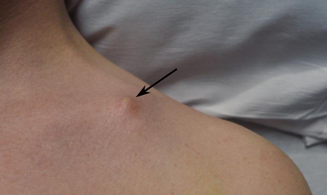
In a clavicle fracture, there is usually an obvious deformity, or “bump,” at the fracture site. Gentle pressure over the break will bring about pain. Although it is rare for a bone fragment to break through the skin, it may push the skin into a “tent” formation.Tenting of skin over clavicle fracture

In a clavicle fracture, the broken ends of the bone may cause tenting of the skin over the fracture site.

Your doctor will also perform tests to ensure that no nerves or blood vessels were damaged when the fracture occurred.

Imaging Studies

X-rays. X-rays provide images of dense structures, such as bone. Your doctor will order an x-ray to help pinpoint the location of the fracture and to learn more about the severity of the break.

He or she may also order x-rays of your entire shoulder to check for additional injuries. If other bones are broken, your doctor may order a computerized tomography (CT) scan to see the fractures in better detail.

History and Physical Examination

Left sided displaced clavicle fracture.

The patient may appear with the following signs and symptoms:

  • A patient may cradle the injured extremity with the uninjured arm.
  • A patient may report a snapping or cracking sound when the injury occurs.
  • The shoulder may appear shortened relative to the opposite side and may droop.
  • Swelling, ecchymosis, and tenderness may be noted over the clavicle.
  • Abrasion over the clavicle may be noted, suggesting that the fracture was from a direct mechanism.
  • Crepitus from the fracture ends rubbing against each other may be noted with gentle manipulation.
  • Difficulty breathing or diminished breath sounds on the affected side may indicate a pulmonary injury, such as a pneumothorax.
  • Palpation of the scapula and ribs may reveal a concomitant injury.
  • Tenting and blanching of the skin at the fracture site may indicate an impending open fracture, which most often requires surgical stabilization.
  • Nonuse of the arm on the affected side is a neonatal presentation.
  • Associated distal nerve dysfunction indicates a brachial plexus injury.
  • Decreased pulses may indicate a subclavian artery injury.
  • Venous stasis, discoloration, and swelling indicate a subclavian venous injury.

Diagnostic Procedures and Differential Diagnosis

Diagnose can often be made by a client’s history and physical examination. 

The differential diagnosis of a clavicle fracture includes acromioclavicular joint injury, rib fracture, scapular fracture, shoulder dislocation, rotator cuff injury, and sternoclavicular joint injury.

Possible complications of clavicle fractures must also be fully evaluated, including pneumothorax, brachial plexus injury, and subclavian vessel injury.

Laboratory studies are ordered in clavicle fractures according to the severity of trauma. With a suspected vascular injury, obtain a complete blood count (CBC) to check the hemoglobin and hematocrit values. If a pulmonary injury is suspected or identified, perform an arterial blood gas (ABG) test and obtain an expiration posteroanterior (PA) chest film. Other imaging studies that can be used in the assessment of a clavicle fracture include the following:

  • Radiography of the clavicle and shoulder
  • Computed tomography (CT) scanning with 3-dimensional (3-D) reconstruction
  • Arteriography
  • Ultrasonography

Laboratory studies are ordered in clavicle fractures according to the severity of trauma. With a suspected vascular injury, obtain a complete blood count (CBC) to check the hemoglobin and hematocrit values. If a pulmonary injury is suspected or identified, perform an arterial blood gas (ABG) test and obtain an expiration posteroanterior (PA) chest film. Other imaging studies that can be used in the assessment of a clavicle fracture include the following:

  • Radiography of the clavicle and shoulder
  • Computed tomography (CT) scanning with 3-dimensional (3-D) reconstruction
  • Arteriography
  • Ultrasonography

Management

Clavicle fracture is managed either surgically or conservatively based upon various factors including the location (mid-shaft, distal, proximal), nature (displaced, undisplaced, comminuted) of the fracture, open VS closed injury, age, and neurovascular compromises.

Traditionally, the management of clavicle fractures has been through conservative management with sling immobilization and subsequent rehabilitation. This continues to provide satisfactory results for undisplaced fractures but conservative management of displaced mid-shaft clavicle fractures results in increased rates of re-injury, increased return times to sport and suboptimal shoulder function, secondary to clavicular mal-union and shortening, with resultant thoracoscapular dyskinesia. Similarly, conservative management of displaced lateral fractures in the athletic patient has been shown to result in high rates of non-union and subsequent impairment of shoulder function.

So for the athletic individual, operative intervention is routinely performed for displaced lateral fractures and is recommended for mid-shaft fractures that are completely displaced, shortened >2 cm or comminuted.

REQUEST AN APPOINTMENT OR BOOK A CONSULANT – Sargam.dange.18@gmail.com

Surgical Treatment

The chief goal of this treatment is to achieve a healed clavicular strut in a normal anatomical position as possible.

Indications for operative treatment of clavicular fractures are;

  1. Severe displacement caused by comminution with resultant angulation and tenting of the skin severe enough to threaten its integrity and that fails to respond to closed reduction.
  2. Symptomatic non-union like shoulder girdle dysfunction neurovascular compromise.
  3. Neurovascular injury or compromise that is progressive or that fails to revere after the closed reduction of the fracture.
  4. Open fracture.
  5. Type II distal clavicular fracture (displaced).
  6. Multiple traumas, when mobility of the patients is desirable and closed methods of immobilization are impractical or possible.
  7. Floating shoulder.
  8. Inability to tolerate closed immobilization such as neurological problems of Parkinsonism, seizure disorders.
  9. Cosmetic reasons.
  10. Relative indications include shortening of more than 15 to 20mm and displacement more than the width of the clavicle.

Surgical procedure includes:

  • Internal fixation with plates and screws. ( most common)
  • Intramedullary (IM) fixation.

The routine removal of metalwork was recommended for IM Nail but not for plate fixation in midshaft displaced fractures. Whereas, in displaced lateral clavicle fracture routine removal of metalwork was performed for ‘hook’ plate fixation, screw fixation, cerclage wire fixation, and tension band wire fixation but not for ‘non-ACJ-spanning’ plate fixation and suture fixation. These fixation methods are necessary for lateral clavicle fracture as it involves acromioclavicular joint and various ligaments that may become injured during fracture.

Physical Therapy/Rehabilitation

The primary goal of rehabilitation is to improve and restore the function of the shoulder for activities of daily living, vocational, and sports activities. Rehabilitation protocol may vary slightly in first few weeks based upon the primary treatment approach i.e. conservative vs surgical.

Rehabilitation for Conservative Management

Fracture healing may take more time in nonoperative treatment. Conservatively treated fractures of the clavicular midshaft usually unite between 18 and 28 weeks after the injury. So regular follow- up needs to be done to check whether the fracture site is properly unioned or not. So rehabilitation protocol may also vary based upon individual co-morbidities.

In first few weeks ( 2-4 weeks), POLICE principle can be used in acute undisplaced clavicle fracture which is further explained below in context of clavicle fracture.

Protection:

Patient’s shoulder is immobilized in a sling or figure-of-eight brace until the clinical union is achieved. A figure-of-eight brace is often thought to prevent or reduce secondary fracture shortening during the time of fracture healing. But it is associated with more discomfort and pain including nerve compression with temporary brachial plexus palsies and restriction of venous blood return. And studies concluded that there are no differences between these two techniques regarding healing time and the rate of nonunion for treating clavicle fractures. So a sling is usually used and immobilization in internal rotation is usually recommended for 2-4 weeks. Wear the sling during the day, except for exercises and personal hygiene.Patient’s choice to wear at night or not but they need to be cautious.

During forceful coughing, sneezing also patients need to take caution (as respiratory excursions may cause clavicle movement) by avoiding it as much as possible and also learning active-assisted coughing techniques if necessary.

Optimal loading

Therapy/Advice for 1-2 weeks Post injury:

  • Use of arm sling as mentioned above ( need to use most of the times).
  • Self-mobilisation of the elbow and wrist out of the sling is required several times a day to avoid stiffening of the elbow and wrist.
  • Do not lift your elbow above shoulder height as this may be painful.
  • The range of motion of the shoulder should usually be limited to pendulum exercises for the first 1-2 weeks.
  • Correct postural habits and neck ROM are taught.

Therapy/Advice for 3-6 weeks Post -injury:

  • Decrease the use of arm sling (use during non-dependent position).
  • Begin normal light daily activities with the arm and shoulder.
  • Shoulder active-assisted to active range of motion in a single plane with no more above than 90 degrees is recommended within the first 6 weeks.
  • Scapular mobilization exercises are addressed.
  • Isometric exercises of Shoulder with tolerable resistance is started within 4-6 weeks.
  • Avoid heavy lifting for the full 6 weeks.
  • Gradual progression of cardiovascular endurance training can be started using brisk walking and static bycycle.

Therapy/Advice for 6-12 weeks Post-injury:

  • Free active and active assisted range of motion of shoulder in all planes is usually allowed after 6 weeks with passive ROM as tolerable.
  • Progressive resistive exercises (isotonic) for scapular stabilizers, biceps, triceps and rotator cuff are prescribed after 6 weeks.
  • Weight-bearing should be avoided until clinical fracture.
  • Sporting activities and work, demanding weight-bearing and the use of the arm, are usually suspended until the patient is free of pain with radiographic signs of progressing fracture consolidation, usually after 6-12 weeks.

Therapy/Advice for 12 weeks and beyond:

Start a more aggressive strengthening program, cardiovascular endurance training as tolerated, and progressive sports- specific training are addressed.

  • Return to specific sports is determined by the physical therapist through functional testing specific to the patient’s demands according to which progressive sport-specific training is planned.
  • Advance activities including muscle endurance activities (upper body ergometer) and cardiovascular endurance exercises (treadmill, cycling) can be prescribed.
  • Contact sports should be avoided for 3-4 months. Return to full contact sports requires the athlete should demonstrate radiographic evidence of bony healing, no tenderness to palpation, a full range of motion, and normal shoulder strength.

Rehabilitation After Postoperative Treatment

  • Primary open reduction and internal fixation with plate ( locking compression plate) and screws of middle third clavicle fractures provides a more rigid fixation and allow immediate post-surgical mobilzation. Surgical management help bone healing faster than that of conservative treatment. So the duration of immobilization is shorter compare to conservative treatment and mobilization and strengthening exercises can be prescribed in earlier than that of conservative management. A similar progression of exercise can be prescribed as of conservative treatment but progression can be made earlier.

Treatment

Nonsurgical Treatment

If the broken ends of the bones have not significantly shifted out of place, you may not need surgery. Most broken collarbones can heal without surgery.

Nonsurgical treatment may include:

  • Arm support. A simple arm sling is usually used for comfort immediately after the break and to keep your arm and shoulder in position while the injury heals.
  • Medication. Pain medication, including acetaminophen, can help relieve pain as the fracture heals.
  • Physical therapy. Although there will be some pain, it is important to maintain arm motion to prevent stiffness. Often, patients will begin doing exercises for elbow motion immediately after the injury.

    After a clavicle fracture, it is common to lose some shoulder and arm strength. Once the bone begins to heal, your pain will decrease and your doctor may start gentle shoulder exercises. These exercises will help prevent stiffness and weakness. More strenuous exercises will be started gradually once the fracture is completely healed.

Follow-up care. You will need to see your doctor regularly until your fracture heals. During these visits, he or will take x-rays to make sure the bone is healing in a good position. After the bone has healed, you will be able to gradually return to your normal activities.

Complications. In some cases, a clavicle fracture can move out of place before it heals. It is important to follow up with your doctor as scheduled to make sure the bone stays in position.

If the fracture fragments do move out of place and the bones heal in that position, it is called a “malunion.” Treatment for this is determined by how far out of place the bones are and how much this affects your arm movement.

A large bump over the fracture site may develop as the fracture heals. This usually gets smaller over time, but a small bump may remain permanently.

Surgical Treatment

If the broken ends of the bones have significantly shifted out of place, your doctor may recommend surgery.

Surgery typically involves putting the broken pieces of bone back into position and preventing them from moving out of place until they are healed. This can improve shoulder strength when you have recovered.

Open reduction and internal fixation. This is the procedure most often used to treat clavicle fractures. During the procedure, the bone fragments are first repositioned (reduced) into their normal alignment. The pieces of bone are then held in place with special metal hardware.

Common methods of internal fixation include:

  • Plates and screws. After being repositioned into their normal alignment, the bone fragments are held in place with special screws and metal plates attached to the outer surface of the bone.

    After surgery, you may notice a small patch of numb skin below the incision. This numbness will become less noticeable with time. Because the clavicle lies directly under the skin, you may be able to feel the plate through your skin.

    Plates and screws are not routinely removed after the bone has healed, unless they are causing discomfort. Problems with the hardware are not common, but some patients find that seatbelts and backpacks can irritate the collarbone area. If this happens, the hardware can be removed after the fracture has healed.

Internal fixation of clavicle fracture

(Left) X-ray shows a displaced clavicle fracture (arrow). (Right) The pieces of bone have been realigned and held in place with plates and screws.

  • Pins or screws. Pins or screws can also be used to hold the fracture in good position after the bone ends have been put back in place. The incisions for pin or screw placement are usually smaller than those used for plates.

    Pins or screws often irritate the skin where they have been inserted and are usually removed once the fracture has healed.

Internal fixation of clavicle fracture (Left) X-ray shows a severely displaced clavicle fracture (arrow). (Right) Here, a single screw has been used to repair the fracture. Reproduced from Eichinger JK, Balog TP, Grassbaugh JA: Intramedullary fixation of clavicle fractures: anatomy, indications, advantages, and disadvantages. J Am Acad Orthop Surg 2016; 24(7): 455-464.

Pain management. After surgery, you will feel some pain.This is a natural part of the healing process. Many patients find that using ice and simple, non-prescription medications for pain relief are all that is needed to relieve pain.

If your pain is severe, your doctor may suggest a prescription-strength medication, such as an opioid, for a few days.

Be aware that although opioids help relieve pain after surgery, they are a narcotic and can be addictive. Opioid dependency and overdose has become a critical public health issue. For this reason, opioids are typically prescribed for a short period of time. It is important to use opioids only as directed by your doctor. As soon as your pain begins to improve, stop taking opioids.

Rehabilitation. Specific exercises will help restore movement and strengthen your shoulder. Your doctor may provide you with a home therapy plan or suggest that you work with a physical therapist.

Therapy programs typically start with gentle motion exercises. Your doctor will gradually add strengthening exercises to your program as your fracture heals.

Although it is a slow process, following your physical therapy plan is an important factor in returning to all the activities you enjoy.

Complications. There are risks associated with any type of surgery. These include:

  • Infection
  • Bleeding
  • Problems with wound healing
  • Pain
  • Blood clots
  • Damage to blood vessels or nerves
  • Reaction to anesthesia

Risks that are specific to surgery for clavicle fractures include:

  • Difficulty with bone healing
  • Lung injury
  • Hardware irritation

Patients who smoke or use tobacco products, have diabetes, or are elderly are at a higher risk for complications both during and after surgery. They are also more likely to have problems with wound and bone healing.

Before your surgery, your doctor will discuss each of the risks with you and will take specific measures to avoid complications.

REQUEST AN APPOINTMENT OR BOOK A CONSULANT – Sargam.dange.18@gmail.com

post fracture physiotherapy management

REQUEST AN APPOINTMENT OR BOOK A CONSULANT – Sargam.dange.18@gmail.com

A fracture is a discontinuity in a bone (or cartilage) resulting from mechanical forces that exceed the bone’s ability to withstand them.

Most commonly fractures occur in the setting of a normal bone with acute overwhelming force, usually in the setting of trauma. Fractures can also occur, however, in a variety of other settings.

  1. The entire skeleton may be weak due to metabolic (e.g. osteoporosis) or less frequently genetic abnormalities (e.g. osteogenesis imperfecta) and thus prone to fractures from forces that would be insufficient to cause fractures in normal bones. These are known as insufficiency fractures.
  2. The protracted chronic application of abnormal stresses (e.g. running)  can result in the accumulation of microfractures faster than the body can heal, eventually resulting in macroscopic failure. These are termed fatigue fractures. Nb. Together, insufficiency and fatigue fractures are often grouped together as stress fractures.
  3. The bone may have a lesion that focally weakens a bone (e.g. metastasis, or bone cyst). These are known as pathological fractures.

Pathophysiology Of Bone Healing

The pathophysiological sequence of events that occur following a fracture can be divided into three main phases:

  1. Inflammatory
  2. Reparative
  3. Remodeling

Inflammatory Phase

Immediately at the time of fracture, the space between fracture ends is filled with blood-forming a hematoma.

  • Stops additional bleeding; provides structural and biochemical support for the influx of inflammatory cells, fibroblasts, chondroblasts and the ingrowth of capillaries

This process takes approximately a week, forming a primary callus which is non-mineralized and not readily visible on radiography

Reparative Phase

Over the next few weeks, this primary callus is transformed into a bony callus by the activation of osteoprogenitor cells. These cells lay down woven bone which stabilises the fracture site.

Remodeling Phase

This phase lasts many months, maybe years, and represents the gradual formation of compact cortical bone with greater biomechanical properties and allows for the reduction of the width of the callus. Remodeling can result in almost perfect healing, however, particularly if the alignment is not perfect, a residual deformity will remain.

Clinical Features of Fracture

Clinical features vary depending on the cause and nature of the injury and range from unconsciousness to the patient being able to use the limb, although complaining of pain. These features are :

  • Pain (Image at arm following a boxing injury, painful)
  • Deformity
  • Oedema
  • Loss of function
  • Muscle spasm
  • Muscle atrophy
  • Abnormal movement
  • Limitation of joint motion
  • Shock

REQUEST AN APPOINTMENT OR BOOK A CONSULANT – Sargam.dange.18@gmail.com

Treatment and Prognosis

The basics of fracture healing rely on alignment and immobilisation.

Alignment may or may not be necessary depending on the degree of displacement, the importance of correct alignment (e.g. index finger vs rib), and the patient (e.g. professional athlete vs debilitated elderly).

Immobilisation can be achieved in a variety of ways depending on the location, morphology of the fracture, and device of fixation

  • None (e.g. most rib fractures)
  • Sling (e.g. many clavicular fractures)
  • Cast (e.g. many forearm fractures)
  • Internal fixation (e.g. most hip fractures)
  • External fixation

Fixation Devices

Stress sharing devices

It allows micromotion between the two fractured sites and partial transmission of load, so promote secondary bone healing with callus formation, which is a relatively rapid bone healing.

For example; intramedullary nail, casts, rods.

Stress Shielding Devices

The stress at the fracture site is transmitted through the shielding device, there is no motion at the fracture site so promote primary bone healing without callus formation, which is slower than the healing with callus formation.

For example; compression plate.

Role of Physiotherapy

Crutches Walking.png

The physiotherapist’s role is to identify the cause of the problem and to select the appropriate procedure to alleviate or eliminate the cause of the loss of movement. Examples of early treatment include

  • A physical therapist may instruct the client how to walk with an assistive device, like a cane or crutches. This includes how to use the device to walk up and down stairs or to get into and out of a car. Learning a new skill takes practice, so be sure to allow client practice using your device while they are with you.
  • After a lower extremity fracture there may limit the amount of weight client can put on the leg. Help the client understand weight bearing restrictions and teach how to move about while still maintaining these restrictions.
  • If the fracture is in the arm, you as the physical therapist may teach you how to apply and remove the sling

Doing an assessment for the patient is necessary also doing The problem-oriented medical record (POMR) system ( is based on a data collection system that incorporates the acronym SOAP:

  • Subjective – any information given to you by the patient: allergies, past medical history, past surgical history, family history, social history (living arrangements, social conditions, employment, medication), review of systems .
  • Objective – all information obtained through observation or testing, e.g. range of joint movement, muscle strength .
  • Analysis – a listing of problems based on what you know from a review of subjective and objective data.
  • Plan – this refers to the plan of treatment).

Also By Using specific exercises, the aim is to reduce any swelling, regain full muscle power and joint movement, and to bring back full function. The treatment will depend very much on the problems identified during your initial assessment, but may include a mixture of the following:

  • Soft tissue massage, particularly to manage Edema and swelling
  • Scar management if the patient had surgery to fix the fracture
  • Ice therapy
  • Stretching exercises to regain joint range of movement
  • Joint manual therapy and mobilizations to assist in regaining joint mobility
  • Structured and progressive strengthening regime
  • Balance and control work and gait (walking) re-education where appropriate
  • Taping to support the injured area/help with swelling management
  • Return to sport preparatory work and advice where required

REQUEST AN APPOINTMENT OR BOOK A CONSULANT – Sargam.dange.18@gmail.com

Fracture

REQUEST AN APPOINTMENT OR BOOK A CONSULANT – Sargam.dange.18@gmail.com

A fracture is a broken bone. It can range from a thin crack to a complete break. Bone can fracture crosswise, lengthwise, in several places, or into many pieces. Most fractures happen when a bone is impacted by more force or pressure than it can support.

A bone fracture is a medical condition where the continuity of the bone is broken.If you suspect you have a fracture, seek medical help immediately.

A significant percentage of bone fractures occur because of high force impact or stress.However, a fracture may also be the result of some medical conditions which weaken the bones, for example osteoporosis, some cancers, or osteogenesis imperfecta (also known as brittle bone diseases).A fracture caused by a medical condition is known as a pathological fracture.

Types of bone fracture-

There is a range of fracture types, including:

  • Avulsion fracture – a muscle or ligament pulls on the bone, fracturing it.
  • Comminuted fracture – the bone is shattered into many pieces.
  • Compression (crush) fracture – generally occurs in the spongy bone in the spine. For example, the front portion of a vertebra in the spine may collapse due to osteoporosis.
  • Fracture dislocation – a joint becomes dislocated, and one of the bones of the joint has a fracture.
  • Greenstick fracture – the bone partly fractures on one side, but does not break completely because the rest of the bone can bend. This is more common among children, whose bones are softer and more elastic.
  • Hairline fracture – a partial fracture of the bone. Sometimes this type of fracture is harder to detect with routine xrays.
  • Impacted fracture – when the bone is fractured, one fragment of bone goes into another.
  • Intraarticular fracture – where the break extends into the surface of a joint
  • Longitudinal fracture – the break is along the length of the bone.
  • Oblique fracture – a fracture that is diagonal to a bone’s long axis.
  • Pathological fracture – when an underlying disease or condition has already weakened the bone, resulting in a fracture (bone fracture caused by an underlying disease/condition that weakened the bone).
  • Spiral fracture – a fracture where at least one part of the bone has been twisted.
  • Stress fracture – more common among athletes. A bone breaks because of repeated stresses and strains.
  • Torus (buckle) fracture – bone deforms but does not crack. More common in children. It is painful but stable.
  • Transverse fracture – a straight break right across a bone.

Closed vs. open

A closed fracture is also called a simple fracture. In a closed fracture, the broken bone doesn’t break your skin.

An open fracture is also called a compound fracture. In an open fracture, the ends of the broken bone tear your skin. When your bone and other internal tissues are exposed, it puts you at higher risk of infection.

Incomplete vs. complete

In an incomplete fracture, your bone doesn’t break completely. In other words, it cracks without breaking all the way through. Types of incomplete fracture include:

  • hairline fracture, in which your bone is broken in a thin crack
  • greenstick fracture, in which your bone is broken on one side, while the other side is bent
  • buckle or torus fracture, in which your bone is broken on one side and a bump or raised buckle develops on the other side

In a complete fracture, your bone breaks completely. It’s snapped or crushed into two or more pieces. Types of complete fracture include:

  • single fracture, in which your bone is broken in one place into two pieces
  • comminuted fracture, in which your bone is broken or crushed into three or more pieces
  • compression fracture, in which your bone collapses under pressure
  • nondisplaced fracture, in which your bone breaks into pieces that stay in their normal alignment
  • displaced fracture, in which your bone breaks into pieces that move out of their normal alignment
  • segmental fracture, in which your bone is broken in two places in a way that leaves at least one segment floating and unattached

Incomplete fractures are more common in children. Their bones are softer than those of adults. As a result, they’re more likely to bend than break. Complete fractures can happen at any age.

causes

Most fractures are caused by a bad fall or automobile accident. Healthy bones are extremely tough and resilient and can withstand surprisingly powerful impacts. As people age, two factors make their risk of fractures greater: Weaker bones and a greater risk of falling.

Children, who tend to have more physically active lifestyles than adults, are also prone to fractures.

People with underlying illnesses and conditions that may weaken their bones have a higher risk of fractures. Examples include osteoporosis, infection, or a tumor. As mentioned earlier, this type of fracture is known as a pathological fracture.

Stress fractures, which result from repeated stresses and strains, commonly found among professional sports people, are also common causes of fractures.

Fast facts on fractures

Here are some key points about fractures. More detail and supporting information is in the main article.

  • Most bone fractures are caused by falls and accidents.
  • Bone fractures caused by disease are referred to as pathological fractures.
  • A compound fracture is one that also causes injury to the overlying skin.
  • There are a number of different types of fractures, including avulsion, comminuted, and hairline fractures.
  • Bone healing is a natural process, treatment revolves around giving the bone optimum conditions to heal itself.

Symptoms

The signs and symptoms of a fracture vary according to which bone is affected, the patient’s age and general health, as well as the severity of the injury. However, they often include some of the following:

  • pain
  • swelling
  • bruising
  • discolored skin around the affected area
  • angulation – the affected area may be bent at an unusual angle
  • the patient is unable to put weight on the injured area
  • the patient cannot move the affected area
  • the affected bone or joint may have a grating sensation
  • if it is an open fracture, there may be bleeding

When a large bone is affected, such as the pelvis or femur:

  • the sufferer may look pale and clammy
  • there may be dizziness (feeling faint)
  • feelings of sickness and nausea.

If possible, do not move a person with a broken bone until a healthcare professional is present and can assess the situation and, if required, apply a splint. If the patient is in a dangerous place, such as in the middle of a busy road, one sometimes has to act before the emergency services arrive.

REQUEST AN APPOINTMENT OR BOOK A CONSULANT – Sargam.dange.18@gmail.com

Doctor Examination

Your doctor will do a careful examination to assess your overall condition, as well as the extent of the injury. He or she will talk with you about how the injury occurred, your symptoms, and medical history.

The most common way to evaluate a fracture is with x-rays, which provide clear images of bone. Your doctor will likely use an x-ray to verify the diagnosis. X-rays can show whether a bone is intact or broken. They can also show the type of fracture and exactly where it is located within the bone.

fracture diagnosis

Most fractures are caused by a bad fall or automobile accident. Healthy bones are extremely tough and resilient and can withstand surprisingly powerful impacts. As people age, two factors make their risk of fractures greater: Weaker bones and a greater risk of falling.

Children, who tend to have more physically active lifestyles than adults, are also prone to fractures.

People with underlying illnesses and conditions that may weaken their bones have a higher risk of fractures. Examples include osteoporosis, infection, or a tumor. As mentioned earlier, this type of fracture is known as a pathological fracture.

Stress fractures, which result from repeated stresses and strains, commonly found among professional sports people, are also common causes of fractures.

Complications

Heals in the wrong position – this is known as a malunion; either the fracture heals in the wrong position or it shifts (the fracture itself shifts).

Disruption of bone growth – if a childhood bone fracture affects the growth plate, there is a risk that the normal development of that bone may be affected, raising the risk of a subsequent deformity.

Persistent bone or bone marrow infection – if there is a break in the skin, as may happen with a compound fracture, bacteria can get in and infect the bone or bone marrow, which can become a persistent infection (chronic osteomyelitis).

Patients may need to be hospitalized and treated with antibiotics. Sometimes, surgical drainage and curettage is required.

Bone death (avascular necrosis) – if the bone loses its essential supply of blood it may die.

risk factors

Anyone can be experience a fracture. But you’re more likely to develop one if you have brittle bones, or low bone density. You’re more likely to develop brittle bones if you:

  • are older
  • have osteoporosis
  • have endocrine or intestinal disorders
  • are taking corticosteroids
  • are physically inactive
  • drink alcohol
  • smoke

Diagnosis

Medical intervention focuses on supporting the bone as it heals naturally.

A doctor will carry out a physical examination, identify signs and symptoms, and make a diagnosis.

The patient will be interviewed – or friends, relatives, and witnesses if the patient cannot communicate properly – and asked about circumstances that caused the injury or may have caused it.

Doctors will often order an X-ray. In some cases, an MRI or CT scan may also be ordered.

Bone healing is a natural processTrusted Source which, in most cases, will occur automatically. Fracture treatment is usually aimed at making sure there is the best possible function of the injured part after healing.

Treatment also focuses on providing the injured bone with the best circumstances for optimum healing (immobilization).

For the natural healing process to begin, the ends of the broken bone need to be lined up – this is known as reducing the fracture.

The patient is usually asleep under a general anesthetic when fracture reduction is done. Fracture reduction may be done by manipulation, closed reduction (pulling the bone fragments), or surgery.

Immobilization – as soon as the bones are aligned they must stay aligned while they heal. This may include:

  • Plaster casts or plastic functional braces – these hold the bone in position until it has healed.
  • Metal plates and screws – current procedures may use minimally invasive techniques.
  • Intra-medullary nails – internal metal rods are placed down the center of long bones. Flexible wires may be used in children.
  • External fixators – these may be made of metal or carbon fiber; they have steel pins that go into the bone directly through the skin. They are a type of scaffolding outside the body.

Usually, the fractured bone area is immobilized for 2-8 weeks. The duration depends on which bone is affected and whether there are any complications, such as a blood supply problem or an infection.

How is a fracture treated?

If you’re diagnosed with a fracture, the treatment plan will depend on its type and location.

In general, your doctor will try to put the broken bone pieces back into their proper positions and stabilize them as they heal. It’s important to keep pieces of broken bone immobile until they’re mended. During the healing process, new bone will form around the edges of the broken pieces. If they’re properly aligned and stabilized, the new bone will eventually connect the pieces.

Your doctor may use a cast to stabilize your broken bone. Your cast will likely be made from plaster or fiberglass. It will help keep the injured area stabilized and prevent broken bone pieces from moving while they heal.

In rare cases, you may need traction to stabilize the injured area. Traction stretches the muscles and tendons around your bone. Your doctor will administer it using a system of pulleys and weights positioned in a metal frame over your bed. This system will produce a gentle pulling motion that your doctor can use to stabilize the injured area.

For more complex or compound fractures, you may need surgery. Your doctor may use open reduction, and internal fixation or external fixation to keep your bones from moving.

In open reduction and internal fixation, your doctor will first reposition or “reduce” the pieces of broken bone into their normal alignment. Then they will connect or “fix” the broken bone. This occurs by using screws, metal plates, or both. In some cases, your doctor may insert rods through the center of your bone.

In external fixation, your doctor will put pins or screws into your bone above and below the fracture site. They will connect these pins or screws to a metal stabilizing bar positioned on the outside of your skin. The bar will hold your bone in place as it heals.

Your doctor may also prescribe medication to control pain, fight infection, or manage other symptoms or complications. After the initial treatment stages, they may recommend physical therapy or other strategies to help you regain normal use.

healing

Healing – if a broken bone has been aligned properly and kept immobile, the healing process is usually straightforward.

Osteoclasts (bone cells) absorb old and damaged bone while osteoblasts (other bone cells) are used to create new bone.

Callus is new bone that forms around a fracture. It forms on either side of the fracture and grows toward each end until the fracture gap is filled. Eventually, the excess bone smooths off and the bone is as it was before.

The patient’s age, which bone is affected, the type of fracture, as well as the patient’s general health are all factors which influence how rapidly the bone heals. If the patient smokes regularly, the healing process will take longer.

Physical therapy – after the bone has healed, it may be necessary to restore muscle strength as well as mobility to the affected area. If the fracture occurred near or through a joint, there is a risk of permanent stiffness or arthritis – the individual may not be able to bend that joint as well as before.

Surgery – if there was damage to the skin and soft tissue around the affected bone or joint, plastic surgery may be required.

Delayed unions and non-unions

Non-unions are fractures that fail to heal, while delayed unions are those that take longer to heal.

  • Ultrasound therapy – low-intensity ultrasound is applied to the affected area daily. This has been found to help the fracture heal. Studies in this area are still ongoing.
  • Bone graft – if the fracture does not heal, a natural or synthetic bone is transplanted to stimulate the broken bone.
  • Stem cell therapy – studies are currently underway to see whether stem cells can be used to treat fractures that do not heal.

REQUEST AN APPOINTMENT OR BOOK A CONSULANT – Sargam.dange.18@gmail.com

Introducing concept of psychiatric disorders and their classification

REQUEST AN APPOINTMENT OR BOOK A CONSULANT – Sargam.dange.18@gmail.com

A psychological disorder is a condition characterized by abnormal thoughts, feelings, and behaviors. Psychopathology is the study of psychological disorders, including their symptoms, etiology (i.e., their causes), and treatment. The term psychopathology can also refer to the manifestation of a psychological disorder. Although consensus can be difficult, it is extremely important for mental health professionals to agree on what kinds of thoughts, feelings, and behaviors are truly abnormal in the sense that they genuinely indicate the presence of psychopathology. Certain patterns of behavior and inner experience can easily be labeled as abnormal and clearly signify some kind of psychological disturbance.

The person who washes his hands 40 times per day and the person who claims to hear the voices of demons exhibit behaviors and inner experiences that most would regard as abnormal: beliefs and behaviors that suggest the existence of a psychological disorder. But, consider the nervousness a young man feels when talking to attractive women or the loneliness and longing for home a freshman experiences during her first semester of college—these feelings may not be regularly present, but they fall in the range of normal. So, what kinds of thoughts, feelings, and behaviors represent a true psychological disorder? Psychologists work to distinguish psychological disorders from inner experiences and behaviors that are merely situational, idiosyncratic, or unconventional.

Definition of a Psychological Disorder

Perhaps the simplest approach to conceptualizing psychological disorders is to label behaviors, thoughts, and inner experiences that are atypical, distressful, dysfunctional, and sometimes even dangerous, as signs of a disorder. For example, if you ask a classmate for a date and you are rejected, you probably would feel a little dejected. Such feelings would be normal. If you felt extremely depressed—so much so that you lost interest in activities, had difficulty eating or sleeping, felt utterly worthless, and contemplated suicide—your feelings would be atypical, would deviate from the norm, and could signify the presence of a psychological disorder. Just because something is atypical, however, does not necessarily mean it is disordered.

The American Psychiatric Association (APA) Definition

Many of the features of the harmful dysfunction model are incorporated in a formal definition of psychological disorder developed by the . According to the American Psychiatric Association (APA) (2013), a psychological disorder is a condition that is said to consist of the following:

  • There are significant disturbances in thoughts, feelings, and behaviors. A person must experience inner states (e.g., thoughts and/or feelings) and exhibit behaviors that are clearly disturbed—that is, unusual, but in a negative, self-defeating way. Often, such disturbances are troubling to those around the individual who experiences them. For example, an individual who is uncontrollably preoccupied by thoughts of germs spends hours each day bathing, has inner experiences, and displays behaviors that most would consider atypical and negative (disturbed) and that would likely be troubling to family members.
  • The disturbances reflect some kind of biological, psychological, or developmental dysfunction. Disturbed patterns of inner experiences and behaviors should reflect some flaw (dysfunction) in the internal biological, psychological, and developmental mechanisms that lead to normal, healthy psychological functioning. For example, the hallucinations observed in schizophrenia could be a sign of brain abnormalities.
  • The disturbances lead to significant distress or disability in one’s life. A person’s inner experiences and behaviors are considered to reflect a psychological disorder if they cause the person considerable distress, or greatly impair his ability to function as a normal individual (often referred to as functional impairment, or occupational and social impairment). As an illustration, a person’s fear of social situations might be so distressing that it causes the person to avoid all social situations (e.g., preventing that person from being able to attend class or apply for a job).
  • The disturbances do not reflect expected or culturally approved responses to certain events. Disturbances in thoughts, feelings, and behaviors must be socially unacceptable responses to certain events that often happen in life. For example, it is perfectly natural (and expected) that a person would experience great sadness and might wish to be left alone following the death of a close family member. Because such reactions are in some ways culturally expected, the individual would not be assumed to signify a mental disorder.

REQUEST AN APPOINTMENT OR BOOK A CONSULANT – Sargam.dange.18@gmail.com

unites of classification-

  • Diseases?
  • Disorders?
  • Syndromes?

In psychiatry most of the disorders or diseases diagnosed are syndromes syndromes syndrome a collections of symptoms that tend to appear collections of symptoms that tend to appear together and that seem to have a characteristic course and outcome Purposes of diagnosis in psychiatry help to simplify your thinking and reduce the complexity of clinical phenomena the complexity of clinical phenomena facilitate communication between clinicians (concisely summarizes information for all other clinicians)information for all other clinicians)help to predict the outcome of the disorder decide on an appropriate treatment assist in the search for pathophysiology and etiology.

Neurosis and psychosis

The traditional division between neurosis and psychosis that was evident in ICD-9 (although deliberately left without any attempt to define these concepts) has not been used in ICD-10. However, the term “neurotic” is still retained for occasional use and occurs, for instance, in the heading of a major group (or block) of disorders F40-F48, “Neurotic, stress-related and somatoform disorders”. Except for depressive neurosis, most of the disorders regarded as neuroses by those who use the concept are to be found in this block,and the remainder are in the subsequent blocks. Instead of following the neurotic-psychotic dichotomy, the disorders are now arranged in groups according to major common themes or descriptive likenesses, which makes for increased convenience of use. For instance, cyclothymia (F34.0) is in the block F30-F39, Mood [affective] disorders, rather than in F60-F69, Disorders of adult personality and behaviour; similarly, all disorders associated with the use of psychoactive substances are grouped together in F10-F19, regardless of their severity. “Psychotic” has been retained as a convenient descriptive term, particularly in F23, Acute and transient psychotic disorders. Its use does not involve assumptions about psychodynamic mechanisms, but simply indicates the presence of hallucinations, delusions, or a limited number of severe abnormalities of behaviour, such as gross excitement and overactivity, marked psychomotor retardation, and catatonic behaviour.

Psychogenic and psychosomatic

The term “psychogenic” has not been used in the titles of categories, in view of its different meanings in different languages and psychiatric traditions. It still occurs occasionally in the text, and should be taken to indicate that the diagnostician regards obvious life events or difficulties as playing an important role in the genesis of the disorder. “Psychosomatic” is not used for similar reasons and also because use of this term might be taken to imply that psychological factors play no role in the occurrence, course and outcome of other diseases that are not so described. Disorders described as psychosomatic in other classifications can be found here in F45.- (somatoform disorders), F50.- (eating disorders), F52.- (sexual dysfunction), and F54.- (psychological or behavioural factors associated with disorders or diseases classified elsewhere). It is particularly important to note category F54.- (category 316 in ICD-9) and to remember to use it for specifying the association of physical disorders, coded elsewhere in ICD-10, with an emotional causation. A common example would be the recording of psychogenic asthma or eczema by means of both F54 from Chapter V(F) and the appropriate code for the physical condition from other chapters in ICD-10

REQUEST AN APPOINTMENT OR BOOK A CONSULANT – Sargam.dange.18@gmail.com

Design a site like this with WordPress.com
Get started