Breast cyst

REQUEST AN APPOINTMENT OR BOOK A CONSULANT – Sargam.dange.18@gmail.com

Breast cysts are fluid-filled sacs that are among the most common breast lumps a woman may have. They may also be a cause of other breast symptoms—from pain to, potentially, nipple discharge.

Many women develop noncancerous changes in their breast tissue. It’s one of the conditions known as fibrocystic breast disease, which includes fluid-filled cysts. A cyst may show up in one or both breasts at the same time and in different parts of the breast. Some cysts are so small that patients don’t feel a lump.

For many patients, breast cysts don’t have symptoms: Only about 7 percent of women have a breast cyst that may be felt with the hand.

Simple fluid-filled cysts form from fluid buildup in the breast glands. Microcysts are tiny and may only be spotted under a microscope. Macrocysts, on the other hand, may span from 1 to 2 inches.

Then there are complex cysts, which are either solid or filled with fluid and solids. A complex breast cyst may require a biopsy and drainage to check the cells, a procedure designed to determine whether the cyst is cancerous. On rare occasions, complex cysts are cancerous or increase the risk of breast cancer later.

Symptoms

Breast cysts may be found in one or both breasts. Signs and symptoms of a breast cyst include:

  • A smooth, easily movable round or oval lump that may have smooth edges — which typically, though not always, indicates it’s benign
  • Nipple discharge that may be clear, yellow, straw colored or dark brown
  • Breast pain or tenderness in the area of the breast lump
  • An increase in breast lump size and breast tenderness just before your period
  • A decrease in breast lump size and resolution of other symptoms after your period

Having breast cysts doesn’t increase your risk of breast cancer. But having cysts may make it harder to find new breast lumps or other changes that might need evaluation by your doctor. Your breasts may feel lumpy and painful when you’re menstruating, so it’s important to be familiar with how your breasts feel throughout your menstrual cycle so that you’ll know if something changes.

When to see a doctor

Normal breast tissue often feels lumpy or nodular. But if you feel a new breast lump that doesn’t go away, gets bigger or persists after one or two menstrual cycles, see your doctor right away. Also see your doctor if you have new skin changes on one or both of your breasts.

Causes

Each of your breasts contains lobes of glandular tissue, arranged like petals of a daisy. The lobes are divided into smaller lobules that produce milk during pregnancy and breast-feeding. The supporting tissue that gives the breast its shape is made up of fatty tissue and fibrous connective tissue. Breast cysts develop as a result of fluid accumulation inside the glands in the breasts.

Breast cysts may be defined by their size:

  • Microcysts may be seen during imaging tests, such as mammography or ultrasound, but are too small to feel.
  • Macrocysts are large enough to be felt and can grow to about 1 to 2 inches (2.5 to 5 centimeters) in diameter.

Experts don’t know exactly what causes breast cysts. They may develop as a result of hormonal changes from monthly menstruation.

REQUEST AN APPOINTMENT OR BOOK A CONSULANT – Sargam.dange.18@gmail.com

Diagnosis

Diagnosis of a breast cyst usually includes a breast exam; imaging tests, such as a breast ultrasound or mammogram; and possibly fine-needle aspiration or a breast biopsy.

Breast exam

After discussing your symptoms and health history, your doctor will physically examine the breast lump and check for any other breast abnormalities. Because your doctor can’t tell from a clinical breast exam alone whether a breast lump is a cyst, you’ll need another test. This is usually either an imaging test or fine-needle aspiration.

Imaging tests

Needed tests may include:

  • Mammography. Large cysts and clusters of small cysts can usually be seen with mammography. But microcysts can be difficult or impossible to see on a mammogram.
  • Breast ultrasound. This test can help your doctor determine whether a breast lump is fluid filled or solid. A fluid-filled area usually indicates a breast cyst. A solid-appearing mass most likely is a noncancerous lump, such as a fibroadenoma, but solid lumps also could be breast cancer.

Your doctor may recommend a biopsy to further evaluate a mass that appears solid. If your doctor can easily feel a breast lump, he or she may skip imaging tests and perform fine-needle aspiration to drain the fluid and collapse the cyst.

Fine-needle aspiration

During a fine-needle aspiration, your doctor inserts a thin needle into the breast lump and attempts to withdraw (aspirate) fluid. Often, fine-needle aspiration is done using ultrasound to guide accurate placement of the needle. If fluid comes out and the breast lump goes away, your doctor can make a breast cyst diagnosis immediately.

  • If the fluid is not bloody and has a straw-colored appearance and the breast lump disappears, you need no further testing or treatment.
  • If the fluid appears bloody or the breast lump doesn’t disappear, your doctor may send a sample of the fluid for lab testing and refer you to a breast surgeon or to a radiologist — a doctor trained to perform imaging exams and procedures — for follow-up.
  • If no fluid is withdrawn, your doctor will likely recommend an imaging test, such as a diagnostic mammogram or ultrasound. The lack of fluid or a breast lump that doesn’t disappear after aspiration suggests that the breast lump — or at least a portion of it — is solid. A sample of the tissue may be collected to check for cancer.

Treatment

No treatment is necessary for simple breast cysts — those that are fluid filled and don’t cause any symptoms — that are confirmed on breast ultrasound or after a fine-needle aspiration. Many cysts will disappear with no treatment. If a cyst persists, feels firmer or you notice skin changes on the skin over the cyst, follow up with your doctor.

Fine-needle aspiration

Fine-needle aspiration may be used to diagnose and treat a breast cyst if all the fluid can be removed from the cyst during the procedure, and then your breast lump disappears and your symptoms resolve.

For some breast cysts, however, you may need to have fluid drained more than once. Recurrent or new cysts are common. If a breast cyst persists through two to three menstrual cycles and grows larger, see your doctor for further evaluation.

Hormone use

Using birth control pills (oral contraceptives) to regulate your menstrual cycles may help reduce the recurrence of breast cysts. But because of possible significant side effects, birth control pills or other hormone therapy, such as tamoxifen, is usually recommended only for women with severe symptoms. Discontinuing hormone therapy after menopause may also help prevent breast cysts.

Surgery

Surgery to remove a breast cyst is necessary only in unusual circumstances. Surgery may be considered if an uncomfortable breast cyst recurs month after month or if a breast cyst contains blood-tinged fluid or shows other worrisome signs.

REQUEST AN APPOINTMENT OR BOOK A CONSULANT – Sargam.dange.18@gmail.com

Fibroadenoma

REQUEST AN APPOINTMENT OR BOOK A CONSULANT – Sargam.dange.18@gmail.com

Breast fibroadenomas are the most common type of noncancerous (benign) breast tumor. They are often described as round, moveable, painless breast lumps that feel smooth and rubbery. They can be felt during your monthly breast self-exam and will show up on mammograms and ultrasounds.

Fibroadenomas usually occur in premenopausal women. They may grow during pregnancy, breastfeeding, or estrogen therapy.

Although fibroadenomas have a typical feel on physical examination and appearance on breast ultrasound, the only way to ensure a breast lump is a fibroadenoma is through a biopsy. Proven fibroadenomas may not need treatment unless they are large, fast-growing, or cause bothersome symptoms.

A fibroadenoma is a solid breast lump. This breast lump is not cancer. A fibroadenoma happens most often between ages 15 and 35. But it can be found at any age in anyone who has periods.

A fibroadenoma often causes no pain. It can feel firm, smooth and rubbery. It has a round shape. It might feel like a pea in the breast. Or it may feel flat like a coin. When touched, it moves easily within the breast tissue.

Fibroadenomas are common breast lumps. If you have a fibroadenoma, your health care provider may tell you to watch for changes in its size or feel. You may need a biopsy to check the lump or surgery to remove it. Many fibroadenomas need no further treatment.

Symptoms

A fibroadenoma is a solid breast lump that often causes no pain. It is:

  • Round with distinct, smooth borders
  • Easily moved
  • Firm or rubbery

A fibroadenoma often grows slowly. The average size is about 1 inch (2.5 centimeters). A fibroadenoma can get bigger over time. It may be tender or cause soreness a few days before your period. A large fibroadenoma may hurt when you touch it. But most often, this type of breast lump causes no pain.

You can have a single fibroadenoma or more than one fibroadenoma. They can occur in one or both breasts.

Some fibroadenomas shrink over time. Most fibroadenomas in adolescents shrink over many months to a few years. They then disappear. Fibroadenomas may also change shape over time.

Fibroadenomas may get bigger during pregnancy. They might shrink after menopause.

When to see a doctor

Healthy breast tissue often feels lumpy. Make an appointment with your health care provider if you:

  • Find a new breast lump
  • Notice other changes in your breasts
  • Find that a breast lump you had checked in the past has grown or changed in any way

REQUEST AN APPOINTMENT OR BOOK A CONSULANT – Sargam.dange.18@gmail.com

Causes

The cause of fibroadenomas is not known. They might be related to hormones that control your periods.

Less common types of fibroadenomas and related breast lumps may not act the same as typical fibroadenomas. These types of breast lumps include:

  • Complex fibroadenomas. These are fibroadenomas that can get bigger over time. They can press on or displace nearby breast tissue.
  • Giant fibroadenomas. Giant fibroadenomas grow quickly to larger than 2 inches (5 centimeters). They also can press on nearby breast tissue or push it out of place.
  • Phyllodes tumors. Phyllodes tumors and fibroadenomas are made of similar tissues. But under a microscope, phyllodes tumors look different from fibroadenomas. Phyllodes tumors typically have features associated with growing faster. Most phyllodes tumors are benign. This means they are not cancer. But some phyllodes tumors may be cancer. Or they could become cancer. Phyllodes tumors often cause no pain.

Complications

Common fibroadenomas do not affect your risk of breast cancer. But your risk might go up a bit if you have a complex fibroadenoma or a phyllodes tumor.

Diagnosis

A fibroadenoma may first be discovered on physical examination or during a routine screening mammogram. On a mammogram, fibroadenomas appear as round or oval smooth-edged masses. The outline will be clearly defined, not blurry, and will not be invading the adjacent spaces. Sometimes they are accompanied by coarse calcifications (calcium deposits). Fibroadenomas can look like cysts or a well-contained tumor.

If you notice a lump during an exam or if an abnormality is seen on mammography, a breast ultrasound is usually the next step. This is especially true if you have dense breast tissue, which can make a lump more difficult to view.

On ultrasound, a fibroadenoma will be easier to distinguish from other tissue because of the way it responds to sound waves. It will appear as a dark area with a definite outline. It will look homogeneous (looks the same throughout), round or oval, and may have smooth-edged bumps.

Your doctor may also order digital breast tomosynthesis (DBT), a specialized form of mammography that creates 3D images of the breast, and allows a more detailed evaluation when compared to a regular mammogram.

Biopsy

While imaging tests may suggest a fibroadenoma, the conclusive diagnostic test is usually a breast biopsy, a procedure to remove a sample of tissue for examination in a lab.

Different types of biopsy procedures include fine-needle aspiration or core-needle biopsy, both of which use needles to extract tissue, and open biopsy, a surgical procedure in which a cut is made in the skin to extract sample tissue. Which type your healthcare provider chooses will depend on the characteristics of the lump, its location, and other factors. In women at increased risk for breast cancer, your healthcare provider may strongly recommend surgical removal.

As with any biopsy, breast biopsies carry a risk of a false-negative result that is, the test detects no cancer when cancer is actually present). This is because a breast biopsy may only sample one part of a lump and could miss the cancerous portion. For this reason, you should continue routine breast cancer screening even if you have had a benign biopsy in the past.

Fibroadenoma Treatment

It’s important to distinguish between the two types of fibroadenomas when choosing the best treatment options because of the difference in associated breast cancer risk.

Fibroadenomas are classified as either:

  • Simple: Most fibroadenomas are the simple type; they are more common in younger people. There’s usually just one mass in your breast, with a definite border and very uniform cells. A simple fibroadenoma does not raise your risk for breast cancer.
  • Complex: Complex fibroadenomas are less common but become more common as people age. While they may have a definite border, it’s what is inside this kind of fibroadenoma that makes it different. Under a microscope, a complex fibroadenoma will not look organized and uniform like a simple one. There is often evidence of rapidly dividing cells (hyperplasia) that may have an abnormal appearance (atypia). Having a complex fibroadenoma can raise your risk of developing breast cancer,

Aside from fibroadenoma type, other factors may influence treatment choice, such as:

  • Symptoms, especially discomfort and pain
  • Emotional state, such as undue worry about it being cancer

Be open and honest with your healthcare provider about what you’re experiencing so your concerns can be considered when weighing treatment options.

In considering all of this, your healthcare provider will recommend one of the following fibroadenoma treatment options.

Watch and Wait

Since fibroadenomas are not always troublesome and sometimes shrink on their own, just keeping an eye on their progress is the least invasive way to handle them.

Laser Ablation

In your surgeon’s office, an ultrasound-guided laser device is used to destroy the fibroadenoma, leaving behind only a tiny scar, no sutures, and no change in breast shape. You don’t need general anesthesia, and it’s usually an outpatient procedure, which means no hospital stay.

Cryoablation

Cryoablation is a fast, efficient way to freeze a fibroadenoma. In one office visit, the healthcare provider simply freezes the lump so that healthy tissue can take over. This procedure takes less than 30 minutes and results in a tiny scar.

Radiofrequency Ablation (RFA)

This is a way to remove fibroadenomas without resorting to a lumpectomy. Using local anesthesia and ultrasound guidance, a 6- to 8-millimeter (about 1/4 inch to nearly 1/3 inch) cut is made above the fibroadenoma. Through this cut, a surgical wand is inserted and then tipped by a knife that is heated by radiofrequency current. This helps cut through the breast tissue without causing much bleeding. Once it reaches the target, small wires and robotic arms capture the fibroadenoma and extract it.

Vacuum-Assisted Excision

Breast biopsy systems can now be used as a mostly noninvasive way to remove small fibroadenomas. In less than an hour, and under local anesthesia, a 6-millimeter (1/4 inch) cut is made over the fibroadenoma. Then, with ultrasound guidance, a probe is threaded into the lump, which vacuums out sections of the tissue. Recovery is quick and your scar will be quite small.

High-Frequency Focused Ultrasound (HFU)

This is a relatively new treatment that has been used for fibroadenomas and, sometimes, even for breast cancer. HFU appears to cause the selective destruction of deep tissues without damaging surrounding healthy tissue.

Lumpectomy

Surgical removal can be done if you’re worried about a fibroadenoma. Depending on the relative size of the lump and your breast, a lumpectomy may cause a change in your breast’s size or shape. New fibroadenomas may grow in the area of the first lump, so you should know that surgery is not a guarantee that you’ll never have another fibroadenoma. On the other hand, your fibroadenoma can be carefully examined by a pathologist to make sure your diagnosis is correct.

For pain that may be due to a fibroadenoma, over-the-counter pain medications and applied heat are often used.

Complementary and Alternative Medicine (CAM) Concerns

Certain herbal supplements and diets have been touted as effective treatments for fibroadenomas or their symptoms, but few controlled studies have looked into these remedies. Adopting a healthy lifestyle, including regular exercise and eating lots of fruits and vegetables, is always good advice and may help relieve fibroadenoma symptoms. But remember that these interventions shouldn’t replace proper testing.

REQUEST AN APPOINTMENT OR BOOK A CONSULANT – Sargam.dange.18@gmail.com

Mastalgia

REQUEST AN APPOINTMENT OR BOOK A CONSULANT – Sargam.dange.18@gmail.com

Breast pain (mastalgia) can be described as tenderness, throbbing, sharp, stabbing, burning pain or tightness in the breast tissue. The pain may be constant or it may occur only occasionally, and it can occur in men, women and transgender people.

Breast pain can range from mild to severe. It may occur:

  • Just a few days a month, in the two to three days leading up to a menstrual period. This normal, mild-to-moderate pain affects both breasts.
  • A week or longer each month, starting before a period and sometimes continuing through the menstrual cycle. The pain may be moderate or severe, and affects both breasts.
  • Throughout the month, not related to a menstrual cycle.

In men, breast pain is most commonly caused by a condition called “gynecomastia” (guy-nuh-koh-MAS-tee-uh). This refers to an increase in the amount of breast gland tissue that’s caused by an imbalance of the hormones estrogen and testosterone. Gynecomastia can affect one or both breasts, sometimes unevenly.

In transgender women, hormone therapy may cause breast pain. In transgender men, breast pain may be caused by the minimal amount of breast tissue that may remain after a mastectomy.

Most times, breast pain signals a noncancerous (benign) breast condition and rarely indicates breast cancer. Unexplained breast pain that doesn’t go away after one or two menstrual cycles, or that persists after menopause, or breast pain that doesn’t seem to be related to hormone changes needs to be evaluated.

Causes

Mastalgia can be broken down into two separate categories: cyclic and noncyclic. The causes vary, depending on the kind of mastalgia it is.

Cyclic Mastalgia

Cyclic mastalgia is breast pain that’s related to the hormonal variations associated with the menstrual cycle, which affect how your breasts feel over the course of a month.

The pain is often described as a dull, heavy ache, not focused on any specific spot. It affects both breasts and the armpit areas.

Cyclic mastalgia is typically at its worst shortly before the cycle begins, decreasing on the first day of your period and then going away over the next few days. If hormones are the cause, the symptoms should decrease at certain times during your monthly cycle.

Noncyclic Mastalgia

Noncyclic mastalgia is breast pain that doesn’t vary with the menstrual cycle or with hormonal changes. In postmenopausal females, most breast pain is noncyclic, but you can have it before menopause as well.

The pain may be described as sharp or burning and is often felt only in one breast and one particular area of that breast. It’s also known as “trigger zone” breast pain because you can point to where it hurts. It’s most common between the ages of 30 and 50.1

Noncyclical breast pain is often related to:

  • An injury
  • Surgery
  • Infections
  • Breast fibroadenoma, or a breast cyst
  • Inflammatory breast cancer

Large breasts (either from obesity or genetics) may also be a source of noncyclic mastalgia. They can be especially painful if a supportive bra is not worn. This is due to the stretching of Cooper’s ligaments, which are bands of connective tissue that support and shape breast tissue.

Since this type of pain is linked to menstruation, premenopausal females are most likely to experience it. Cyclical breast pain can also be associated with oral contraceptive pills and HRT.

Fibrocystic breast changes, which can cause breast pain, swelling, or thick areas, involve the lobes, ducts, and connective tissue.

Noncyclic Mastalgia

Noncyclic mastalgia is breast pain that doesn’t vary with the menstrual cycle or with hormonal changes.In postmenopausal females, most breast pain is noncyclic, but you can have it before menopause as well.

The pain may be described as sharp or burning and is often felt only in one breast and one particular area of that breast. It’s also known as “trigger zone” breast pain because you can point to where it hurts. It’s most common between the ages of 30 and 50.1

Noncyclical breast pain is often related to:

  • An injury
  • Surgery
  • Infections
  • Breast fibroadenoma, or a breast cyst
  • Inflammatory breast cancer

Large breasts (either from obesity or genetics) may also be a source of noncyclic mastalgia. They can be especially painful if a supportive bra is not worn. This is due to the stretching of Cooper’s ligaments, which are bands of connective tissue that support and shape breast tissue.

Extramammary breast pain

The term “extramammary” means “outside the breast.” Extramammary breast pain feels like it starts in the breast tissue, but its source is actually outside the breast area. Pulling a muscle in the chest, for example, can cause pain in the chest wall or rib cage that spreads (radiates) to the breast. Arthritis that involves the cartilage in the chest, also known as costochondritis, can also cause pain.

When to see a doctor

REQUEST AN APPOINTMENT OR BOOK A CONSULANT – Sargam.dange.18@gmail.com

Make an appointment with your doctor if breast pain:

  • Continues daily for more than a couple of weeks
  • Occurs in one specific area of your breast
  • Seems to be getting worse over time
  • Interferes with daily activities
  • Awakens you from sleep

Breast cancer risk is very low in people whose main symptom is breast pain, but if your doctor recommends an evaluation, it’s important to follow through.

Diagnosis

The diagnosis of mastalgia is most often made based on symptoms and a physical breast exam, though imaging studies may be done to further evaluate abnormalities noted during a physical exam.

The chance of mastalgia being a symptom of breast cancer is quite low, but it’s important to screen women who are at risk.

Breast cancer is often painless, but breast pain can be a symptom of the disease. If you do have breast pain, call your doctor for an appointment right away.

Treatment

The best treatment for your breast pain depends on what is causing it.

For both cyclic and noncyclic pain, reassurance and ruling out breast cancer is often a relief. If treatment is needed, there are effective treatment options.

Home Remedies

Many home remedies have been tried to decrease cyclic and noncyclic pain, sometimes with good results.

They include:

  • Wearing a well-fitting bra: This can help contain painful breasts and keep them from being pulled uncomfortably.
  • Practicing stress relief: Increased stress can affect hormone levels and lead to more discomfort.
  • Exercising: Physical activity decreases estrogen, but it’s difficult to tell from studies if it really decreases mastalgia.
  • Reducing caffeine and eating a low-fat diet: Studies are mixed on how well this works, but it may help some women.

Keeping a journal can also be helpful, especially if you’re trying to see a pattern in your symptoms, which is the best way to determine the kind of breast pain you have.

Medications

Easing pain with Tylenol (acetaminophen) or nonsteroidal anti-inflammatory drugs (NSAIDs) like Advil (ibuprofen) is another option.

If these first-line interventions don’t help, your healthcare provider may suggest prescription medications like Nolvadex (tamoxifen) and Danocrine (danazol). All of these drugs have side effects of their own.

Studies appeared to show the greatest benefit with tamoxifen, but Danocrine is the only medication approved by the Food and Drug Administration (FDA) for treating mastalgia at this time.

REQUEST AN APPOINTMENT OR BOOK A CONSULANT – Sargam.dange.18@gmail.com

Raynaud’s syndrome

REQUEST AN APPOINTMENT OR BOOK A CONSULANT – Sargam.dange.18@gmail.com

Raynaud’s disease causes some areas of the body — such as fingers and toes — to feel numb and cold in response to cold temperatures or stress. In Raynaud’s disease, smaller arteries that supply blood to the skin narrow. This limits blood flow to affected areas, which is called vasospasm.

Other names for this condition are:

  • Raynaud’s phenomenon.
  • Raynaud syndrome.

Raynaud’s phenomenon is a vasospastic disorder that affects the hands triggered by cold or emotional stress resulting in symptoms of pain, pallor progressing to cyanosis, and coolness of the distal extremities. These changes are thought to be due to intimal layer hypertrophy and inflammation; collagen disposition and fibrosis resulting in vessel lumen occlusion. These vascular alterations have been described in the digital arteries, superficial palmar arch and ulnar artery. Angiographic studies have demonstrated little or no collateral circulation distal to the fibrotic vessels. Radiographic findings in the hands may include soft tissue calcifications when the patient has Raynaud’s as part of CREST syndrome, atrophy of the finger tips, acroosteolysis and intercarpal joint space narrowing. The differential diagnosis for acroosteolysis includes: psoriasis, injury (frostbite or thermal burns), neuropathy (diabetes, leprosy, myelomeningocele), collagen vascular disease, hyperparathyroidism, polyvinyl chloride (PVC) exposure, snake/scorpion venom and familial forms (Hadju-Cheney syndrome).

Symptoms

Symptoms of Raynaud’s disease include:

  • Cold fingers or toes.
  • Areas of skin that turn white then blue. Depending on your skin color, these color changes may be harder or easier to see.
  • Numb, prickly feeling or stinging pain upon warming or stress relief.

During an attack of Raynaud’s, affected areas of the skin usually first turn pale. Next, they often change color and feel cold and numb. When the skin warms and blood flow improves, the affected areas may change color again, throb, tingle or swell.

Raynaud’s most commonly affects fingers and toes. But it also can affect other areas of the body, such as nose, lips, ears and even nipples. After warming up, the return of blood flow to the area can take 15 minutes.

When to see a doctor

See your health care provider right away if you have a history of severe Raynaud’s and get a sore or infection in one of your affected fingers or toes.

Causes

Experts don’t fully understand the cause of Raynaud’s attacks. But blood vessels in the hands and feet appear to react too strongly to cold temperatures or stress.

With Raynaud’s, arteries to the fingers and toes narrow when exposed to cold or stress. The narrowed arteries limit blood flow. Over time, these small arteries can thicken slightly and limit blood flow even more.

Cold temperatures are the most likely cause of an attack. Examples are putting hands in cold water, taking something from a freezer or being in cold air. For some people, emotional stress can trigger an episode.

REQUEST AN APPOINTMENT OR BOOK A CONSULANT – Sargam.dange.18@gmail.com

Primary vs. secondary Raynaud’s

There are two main types of the condition.

  • Primary Raynaud’s. Also called Raynaud’s disease, this most common form isn’t the result of another medical condition. It can be so mild that many people with primary Raynaud’s don’t seek treatment. And it can go away on its own.
  • Secondary Raynaud’s. Also called Raynaud’s phenomenon, this form develops because of another health condition. Although secondary Raynaud’s is less common than the primary form, it tends to be more serious.Symptoms of secondary Raynaud’s usually appear around age 40. That’s later than symptoms appear for primary Raynaud’s.

Causes of secondary Raynaud’s include:

  • Connective tissue diseases. Most people who have a rare disease that leads to hardening and scarring of the skin, known as scleroderma, have Raynaud’s. Other diseases that increase the risk of Raynaud’s include lupus, rheumatoid arthritis and Sjogren’s syndrome.
  • Diseases of the arteries. These include a buildup of fatty deposits in blood vessels that feed the heart and a disorder in which the blood vessels of the hands and feet become inflamed. A type of high blood pressure that affects the arteries of the lungs also may cause secondary Raynaud’s.
  • Carpal tunnel syndrome. This condition involves pressure on a major nerve to the hand. The pressure causes numbness and pain in the hand that can make the hand react more to cold temperatures.
  • Repeated actions or vibration. Typing, playing piano or doing movements like that for long periods can cause overuse injuries. So can using vibrating tools, such as jackhammers.
  • Smoking. Smoking narrows blood vessels.
  • Injuries to the hands or feet. Examples include a wrist fracture, surgery or frostbite.
  • Certain medicines. These include beta blockers for high blood pressure, some migraine medicines, attention-deficit/hyperactivity disorder medicines, certain cancer medicines and some cold medicines.

Risk factors

Risk factors for primary Raynaud’s include:

  • Sex. The condition affects more women than men.
  • Age. Although anyone can develop the condition, primary Raynaud’s often begins between the ages of 15 and 30.
  • Climate. The illness also is more common in people who live in colder climates.
  • Family history. Having a parent, sibling or child with the disease appears to increase the risk of primary Raynaud’s.

Risk factors for secondary Raynaud’s include:

  • Certain diseases. These include conditions such as scleroderma and lupus.
  • Certain jobs. These include jobs that cause repeated trauma, such as using tools that vibrate.
  • Certain substances. This includes smoking, taking medicines that affect the blood vessels and being around certain chemicals, such as vinyl chloride.

Complications

If secondary Raynaud’s is severe, reduced blood flow to fingers or toes could cause tissue damage. But that’s rare.

A completely blocked artery can lead to skin sores or dead tissue. This can be difficult to treat. Rarely, very bad untreated instances might require removing the affected part of the body.

Prevention

To help prevent Raynaud’s attacks:

  • Bundle up outdoors. When it’s cold, wear a hat, scarf, socks and boots, and two sets of mittens or gloves. Thermal underwear might help. A coat with cuffs that close around mittens or gloves helps protect the hands from cold air.Wear earmuffs and a face mask if the tip of your nose and your earlobes get too cold.
  • Warm your car. Run your car heater for a few minutes before driving in cold weather.
  • Take care indoors. Wear socks. To take food out of the refrigerator or freezer, wear gloves, mittens or oven mitts. Some people find it helpful to wear mittens and socks to bed during winter.Because air conditioning can cause attacks, set your air conditioner to a warmer temperature. Use drinking glasses that keep hands from feeling cold.

REQUEST AN APPOINTMENT OR BOOK A CONSULANT – Sargam.dange.18@gmail.com

skin tumors

REQUEST AN APPOINTMENT OR BOOK A CONSULANT – Sargam.dange.18@gmail.com

Skin cancer — the abnormal growth of skin cells — most often develops on skin exposed to the sun. But this common form of cancer can also occur on areas of your skin not ordinarily exposed to sunlight.

There are three major types of skin cancer — basal cell carcinoma, squamous cell carcinoma and melanoma.

You can reduce your risk of skin cancer by limiting or avoiding exposure to ultraviolet (UV) radiation. Checking your skin for suspicious changes can help detect skin cancer at its earliest stages. Early detection of skin cancer gives you the greatest chance for successful skin cancer treatment.

Symptoms

Where skin cancer develops

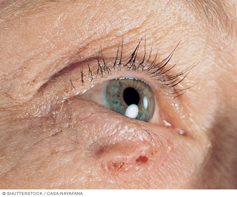
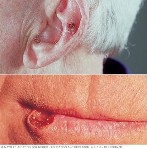
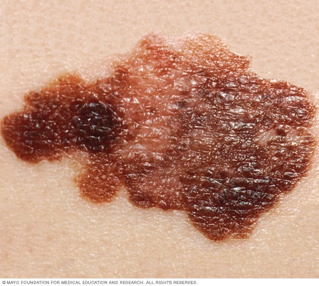
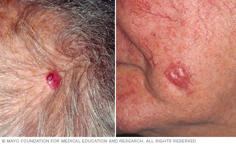
Basal cell carcinoma on white skinBasal cell carcinoma on white skinEnlarge imageSquamous cell carcinoma on the ear and on the lip Squamous cell carcinoma of the skinEnlarge imageMelanomaMelanomaEnlarge imagePhotograph showing Merkel cell carcinoma Merkel cell carcinomaEnlarge image

Skin cancer develops primarily on areas of sun-exposed skin, including the scalp, face, lips, ears, neck, chest, arms and hands, and on the legs in women. But it can also form on areas that rarely see the light of day — your palms, beneath your fingernails or toenails, and your genital area.

Skin cancer affects people of all skin tones, including those with darker complexions. When melanoma occurs in people with dark skin tones, it’s more likely to occur in areas not normally exposed to the sun, such as the palms of the hands and soles of the feet.

Basal cell carcinoma signs and symptoms

Basal cell carcinoma usually occurs in sun-exposed areas of your body, such as your neck or face.

Basal cell carcinoma may appear as:

  • A pearly or waxy bump
  • A flat, flesh-colored or brown scar-like lesion
  • A bleeding or scabbing sore that heals and returns

Squamous cell carcinoma signs and symptoms

Most often, squamous cell carcinoma occurs on sun-exposed areas of your body, such as your face, ears and hands. People with darker skin are more likely to develop squamous cell carcinoma on areas that aren’t often exposed to the sun.

Squamous cell carcinoma may appear as:

  • A firm, red nodule
  • A flat lesion with a scaly, crusted surface

Melanoma signs and symptoms

Melanoma can develop anywhere on your body, in otherwise normal skin or in an existing mole that becomes cancerous. Melanoma most often appears on the face or the trunk of affected men. In women, this type of cancer most often develops on the lower legs. In both men and women, melanoma can occur on skin that hasn’t been exposed to the sun.

Melanoma can affect people of any skin tone. In people with darker skin tones, melanoma tends to occur on the palms or soles, or under the fingernails or toenails.

Melanoma signs include:

  • A large brownish spot with darker speckles
  • A mole that changes in color, size or feel or that bleeds
  • A small lesion with an irregular border and portions that appear red, pink, white, blue or blue-black
  • A painful lesion that itches or burns
  • Dark lesions on your palms, soles, fingertips or toes, or on mucous membranes lining your mouth, nose, vagina or anus

Signs and symptoms of less common skin cancers

Other, less common types of skin cancer include:

  • Kaposi sarcoma. This rare form of skin cancer develops in the skin’s blood vessels and causes red or purple patches on the skin or mucous membranes.Kaposi sarcoma mainly occurs in people with weakened immune systems, such as people with AIDS, and in people taking medications that suppress their natural immunity, such as people who’ve undergone organ transplants.Other people with an increased risk of Kaposi sarcoma include young men living in Africa or older men of Italian or Eastern European Jewish heritage.
  • Merkel cell carcinoma. Merkel cell carcinoma causes firm, shiny nodules that occur on or just beneath the skin and in hair follicles. Merkel cell carcinoma is most often found on the head, neck and trunk.
  • Sebaceous gland carcinoma. This uncommon and aggressive cancer originates in the oil glands in the skin. Sebaceous gland carcinomas — which usually appear as hard, painless nodules — can develop anywhere, but most occur on the eyelid, where they’re frequently mistaken for other eyelid problems.

When to see a doctor

Make an appointment with your doctor if you notice any changes to your skin that worry you. Not all skin changes are caused by skin cancer. Your doctor will investigate your skin changes to determine a cause.

REQUEST AN APPOINTMENT OR BOOK A CONSULANT – Sargam.dange.18@gmail.com

pathophysiology

Although the exact etiology of BCC is unknown, a well-established relationship exists between BCC and the pilosebaceous unit, as tumors are most often discovered on hair-bearing areas.

Many believe that BCCs arise from pluripotential cells in the basal layer of the epidermis or follicular structures. These cells form continuously during life and can form hair, sebaceous glands, and apocrine glands. Tumors usually arise from the epidermis and occasionally arise from the outer root sheath of a hair follicle, specifically from hair follicle stem cells residing just below the sebaceous gland duct in an area called the bulge.

Signaling pathways

The patched/hedgehog intracellular signaling pathway plays a role in both sporadic BCCs and nevoid BCC syndrome (Gorlin syndrome). This pathway influences differentiation of various tissues during fetal development. After embryogenesis, it continues to function in regulation of cell growth and differentiation. Loss of inhibition of this pathway is associated with human malignancy, including BCC.

The hedgehog gene encodes an extracellular protein that binds to a cell membrane receptor complex to start a cascade of cellular events leading to cell proliferation. Of the 3 known human homologues, sonic hedgehog (SHH) protein is the most relevant to BCC. Patched (PTCH) is a protein that is the ligand-binding component of the hedgehog receptor complex in the cell membrane. The other protein member of the receptor complex, smoothened (SMO), is responsible for transducing hedgehog signaling to downstream genes. 

When SHH is present, it binds to PTCH, which then releases and activates SMO. SMO signaling is transduced to the nucleus via Gli. When SHH is absent, PTCH binds to and inhibits SMO. Mutations in the PTCH gene prevent it from binding to SMO, simulating the presence of SHH. The unbound SMO and downstream Gli are constitutively activated, thereby allowing hedgehog signaling to proceed unimpeded. The same pathway may also be activated via mutations in the SMO gene, which also allows unregulated signaling of tumor growth.

How these defects cause tumorigenesis is not fully understood, but most BCCs have abnormalities in either PTCH or SMO genes. Some even consider defects in the hedgehog pathway to be requirements for BCC development.

BCC most commonly develops on sun-exposed areas. Zhang et al reported that ultraviolet (UV)-specific nucleotide changes in PTCH, as well as the tumor suppressor gene TP53, are implicated in the development of early-onset BCC. 

UV-induced mutations in the TP53 tumor suppressor gene, which resides on band 17p13.1, have been found in some cases of BCC.   A germline single-nucleotide polymorphism (SNP) in the TP53 gene, rs78378222, has also been associated with susceptibility to BCC.  In addition, frameshift mutations of the BAX gene (BCL2-associated X protein) have been found in sporadic cases of BCC. A reduction of bcl-2 proteins is observed in the aggressive, infiltrative type of BCC.

REQUEST AN APPOINTMENT OR BOOK A CONSULANT – Sargam.dange.18@gmail.com

Radiation and immunologic origins

Radiation has proven to be tumorigenic by two mechanisms. The first entails the initiations of prolonged cellular proliferation, thereby increasing the likelihood of transcription errors that can lead to cellular transformation. The second mechanism is direct damage of DNA replication, leading to cellular mutation that may activate proto-oncogenes or deactivate tumor suppressor genes.

Immunologically, the mechanism by which prolonged ultraviolet radiation exposure leads to the development of BCC includes suppression of the cutaneous immune system and immunologic unresponsiveness to cutaneous tumors. This local effect includes a decrease in Langerhans cells, dendritic epidermal T cells, and Thy1+ cells. Furthermore, systemic proliferation of suppressor T cells and the release of immunosuppressive factors (eg, tumor necrosis factor-alpha [TNF-alpha], interleukin 1 [IL-1], prostaglandin [PG], interleukin 10 [IL-10]) are believed to be pathogenic to the development of BCC.

DNA mismatch repair proteins

DNA mismatch repair (MMR) proteins are a group of proteins that physiologically stimulate G2 cell cycle checkpoint arrest and apoptosis. Failure of MMR proteins to detect induced DNA damage results in the survival of mutating cells. MMR protein levels have been found to be higher in nonmelanoma skin cancers than in normal skin, and there is also some evidence of MMR dysregulation. 

hysterical neurosis

REQUEST AN APPOINTMENT OR BOOK A CONSULANT – Sargam.dange.18@gmail.com

Hysteria can be defined as a feature of some conditions that involve people experiencing physical symptoms that have a psychological cause.

Hysteria: Disease mainly of women, characterized by lack of control over emotions and acts.
Neurasthenia:   Depression due to exhausted nerve energy.
Neurosis:  A nervous disease, especially a functional disease.
Traumatic neuritis: Inflammation of a nerve caused by an injury.
Malingering:   Feigning illness.

Symptoms of Hysteria

Symptoms that are considered characteristic of “hysteria” include:

  • Blindness
  • Emotional outbursts
  • Hallucinations
  • Histrionic behavior (being overly dramatic or excitable)
  • Increased suggestibility
  • Loss of sensation

Additional symptoms often associated with being in a hysterical state have varied, but include:

  • Being in a sort of trance
  • Developing amnesia
  • Experiencing paralysis
  • Fainting or passing out (syncope)
  • Having epileptic-like seizures
  • Increased pain sensations
  • Rigid or spasming muscles

Diagnosis of Hysteria

In 1980, the American Psychological Association (APA) changed the diagnosis of “hysterical neurosis, conversion type” to “conversion disorder.” Today, someone might be diagnosed with different types of disorders that were historically known as hysteria, including dissociative and somatic disorders.

REQUEST AN APPOINTMENT OR BOOK A CONSULANT – Sargam.dange.18@medicurio

Dissociative Disorders

Dissociative disorders are psychological disorders that involve an interruption in aspects of consciousness, including those related to identity and memory. Disorders in this category include:

  • Dissociative amnesia, which involves forgetting personal information or not being able to recall certain events
  • Dissociative fugue, which involves forgetting personal information combined with changing physical locations, sometimes creating a new identity in the process
  • Dissociative identity disorder, which involves having two or more distinct personalities, each with no memory of what the other has done

Somatic Symptom Disorder

In the DSM-5, symptoms that once existed under the broad umbrella of “hysteria” now fit under what is referred to as somatic symptom disorder.There are several related conditions that can be diagnosed within this category, including:

  • Conversion disorder (functional neurological symptom disorder)
  • Factitious disorder (Munchausen syndrome imposed on one’s self)
  • Illness anxiety disorder (formerly hypochondriasis)

Somatic symptom disorder involves having a significant focus on physical symptoms such as weakness, pain, or shortness of breath. This preoccupation with symptoms results in significant distress and difficulties with normal functioning.

With somatic symptom disorder, the person may or may not have a medical condition. It is important to note that the disorder does not involve faking an illness—whether the person is sick or not, they believe that they are ill. 

Causes of Hysteria

Over time, the theories about what causes hysteria have changed. Although they originally focused on the uterus, we now know that the causes of both dissociative and somatic disorders are often psychological in nature.

Dissociative disorders are generally caused by experiencing some type of trauma. This might include being exposed to childhood abuse that is physical, sexual, or emotional. Being in a natural disaster or being involved in combat can also lead to a dissociative disorder.

Somatic symptom disorder can also be a result of childhood abuse or parental neglect but is also sometimes caused by having extreme anxiety about bodily processes and illness combined with a low threshold for pain.

A phenomenon known as hysterical contagion, a form of social contagion, can also lead groups of people to experience symptoms often associated with hysteria. This phenomenon involves groups of individuals having symptoms of illness that are attributed to a physical or contagious source, but that, in reality, stem from social and psychological influences.

Treatment for Hysteria

Treating hysteria-like symptoms associated with dissociative and somatic symptoms disorders typically includes some type of psychotherapy. Common treatment approaches for these conditions include:

  • Cognitive behavioral therapy (CBT)
  • Dialectical behavioral therapy (DBT)
  • Eye movement desensitization and reprocessing (EMDR)
  • Mindfulness-based therapy

In some cases, medications may also be used to help reduce symptoms. For example, amitriptyline, selective serotonin reuptake inhibitors (SSRIs), and St. John’s wort are known for effectively treating somatic symptom disorder.

Coping With Hysteria

If you or a loved one experience symptoms once associated with hysteria—such as having emotional outbursts, losing sensation, or having hallucinations—seeking the help of a mental health professional is a good step. In the meantime, here are a few tips to help you better cope:

  • Practice mindfulness. Concentrate on the present instead of focusing on yesterday or tomorrow. This can help you stay grounded.
  • Engage in breathing exercises. Relax your feelings of anxiety by breathing in and out in specific patterns.
  • Write in a journal. Get your feelings out and on paper. Put the sources of your stress in black and white, then let them go.
  • Get physically active. Go for a walk or hike, or take your bike for a ride around the neighborhood. Physical activity helps boost mental health while promoting physical health at the same time.
  • Develop a consistent sleep schedule. Give your body the rest it needs to help it better deal with the symptoms and emotions you are experiencing.
  • REQUEST AN APPOINTMENT OR BOOK A CONSULANT – Sargam.dange.18@gmail.com

for consultant or book an online appointment- gmail- sargam.dange.18@gmail.com

behavioural psychology

REQUEST AN APPOINTMENT OR BOOK A CONSULANT – Sargam.dange.18@gmail.com

Behavioral psychology, or behaviorism, is a theory suggesting that environment shapes human behavior. In a most basic sense, behavioral psychology is the study and analysis of observable behavior.

This field of psychology influenced thought heavily throughout the middle of the 20th century. It is still used by mental health professionals today, as its concepts and theories remain relevant in fields like psychotherapy and education

Techniques from Behavioral Psychology

Several concepts in behaviorism are utilized in therapy.

  • Systematic desensitization is used for clients who have a specific phobia, which is characterized by marked fear or anxiety about an object or situation, like an animal or airplanes. Therapy involves applying relaxation or coping techniques as people are gradually exposed to the object or situation.
  • Exposure and response prevention is a strategy that involves exposure to fearful situations, and then not engaging in unhelpful coping strategies. This therapeutic technique is used for obsessive-compulsive disorder (OCD) and other types of anxiety disorders.
  • Token economy reinforces target behavior by giving children and adults symbols or tokens that can be exchanged for something else. It can be used for people with a wide range of mental health issues, as well as in educational settings.
  • Modeling involves clients learning behavior by imitation alone. It’s used in developmental psychology and can be incorporated into clinical use.
  • Applied behavior analysis emerged in the 1960s as a way to modify behavior. It is commonly used for children with an autism spectrum disorder, and is also relevant to fields like education, industrial safety, and criminal behavior.
  • Contingency management involves individuals receiving vouchers for retail goods and services, or the opportunity to win prizes. Often used for patients with substance abuse or related disorders, it typically takes the form of monetary-based reinforcers for drug-negative tests, according to The Psychiatrist.
  • REQUEST AN APPOINTMENT OR BOOK A CONSULANT – Sargam.dange.18@gmail.com

for consultant or book an online appointment- gmail- sargam.dange.18@gmail.com

pyoderma gangrenosum

REQUEST AN APPOINTMENT OR BOOK A CONSULANT – Sargam.dange.18@gmail.com

Pyoderma gangrenosum (pie-o-DUR-muh gang-ruh-NO-sum) is a rare condition that causes large, painful sores (ulcers) to develop on your skin, most often on your legs.

The exact causes of pyoderma gangrenosum are unknown, but it appears to be a disorder of the immune system. People who have certain underlying conditions, such as inflammatory bowel disease or arthritis, are at higher risk of pyoderma gangrenosum.

Pyoderma gangrenosum ulcers can develop quickly. They usually clear up with treatment, but scarring and recurrences are common.

Symptoms

Pyoderma gangrenosum usually starts with a small, red bump on your skin, which may resemble a spider bite. Within days, this bump can develop into a large, painful open sore.

The ulcer usually appears on your legs, but may develop anywhere on your body. Sometimes it appears around surgical sites. If you have several ulcers, they may grow and merge into one larger ulcer.

Causes

The exact cause of pyoderma gangrenosum is unknown. The condition is not infectious or contagious. It’s often associated with autoimmune diseases such as ulcerative colitis, Crohn’s disease and arthritis. And it may have a genetic component.

If you have pyoderma gangrenosum, new skin trauma, such as a cut or puncture wound, may trigger new ulcers.

REQUEST AN APPOINTMENT OR BOOK A CONSULANT – Sargam.dange.18@gmail.com

Risk factors

Certain factors may increase your risk of pyoderma gangrenosum, including:

  • Your age and sex. The condition can affect anyone at any age, though it’s more common between 20 and 50 years of age.
  • Having inflammatory bowel disease. People with a digestive tract disease such as ulcerative colitis or Crohn’s disease are at increased risk of pyoderma gangrenosum.
  • Having arthritis. People with rheumatoid arthritis are at increased risk of pyoderma gangrenosum.
  • Having a blood disorder. People with acute myelogenous leukemia, myelodysplasia or a myeloproliferative disorder are at increased risk of pyoderma gangrenosum.

Complications

Possible complications of pyoderma gangrenosum include infection, scarring, uncontrolled pain, depression and loss of mobility.

Prevention

You can’t totally prevent pyoderma gangrenosum. If you have the condition, try to avoid injuring your skin. Injury or trauma to your skin, including from surgery, can provoke new ulcers to form. It may also help to control any underlying condition that may be causing the ulcers.

Diagnosis

Your doctor will talk with you about your medical history and conduct a physical exam. No test can confirm a diagnosis of pyoderma gangrenosum. But your doctor may order a variety of tests to rule out other conditions that may have similar signs or symptoms. These may include blood tests, a skin biopsy and other tests.

Your doctor may refer you to a specialist in skin conditions (dermatologist).

Treatment

Treatment of pyoderma gangrenosum is aimed at reducing inflammation, controlling pain, promoting wound healing and controlling any underlying disease. Your treatment will depend on several factors, including your health and the number, size, depth and growth rate of your skin ulcers.

Some people respond well to treatment with a combination of pills, creams or injections. Others may need a stay in the hospital or burn treatment center for specialized wound care. Even after successful treatment, it’s common for new wounds to develop.

Medications

  • Corticosteroids. The most common treatment are daily doses of corticosteroids. These drugs may be applied to the skin, injected into the wound or taken by mouth (prednisone). Using corticosteroids for a long time or in high doses may cause serious side effects. Because of this your doctor may prescribe steroid-sparing (nonsteroidal) drugs if you need long-term treatment.
  • Steroid-sparing drugs. An effective nonsteroidal drug is cyclosporine. Other options include mycophenolate (Cellcept), immunoglobulins, dapsone, infliximab (Remicade) and tacrolimus (Protopic), which is a calcineurin inhibitor. Depending on the type of drug used, it may be applied to the wounds, injected or taken by mouth.
  • Pain medication. Depending on the extent of your wounds, you may benefit from pain medication, especially when dressings are being changed.

Wound care

In addition to applying medicine directly to your wounds, your doctor or wound care specialist will cover them with a nonadherent, moist (not wet or dry) dressing and, perhaps, an elasticized wrap. You may be asked to keep the affected area elevated.

Follow your doctor’s instructions regarding wound care. This is especially important because many of the oral medications prescribed for pyoderma gangrenosum suppress your immune system, which increases your risk of infection.

Surgery

Because pyoderma gangrenosum can be made worse by cuts to the skin, surgery to remove dead tissue is not usually considered a good treatment option. Trauma to the skin may worsen existing ulcers or trigger new ones.

If the ulcers on your skin are large and need help with healing, your doctor might suggest a skin graft. In this procedure, the surgeon attaches a piece of skin or synthetic skin over the open sores. This is attempted only after the wound inflammation has gone and the ulcer has started healing.

for consultant or book an online appointment- gmail- sargam.dange.18@gmail.com

REQUEST AN APPOINTMENT OR BOOK A CONSULANT – Sargam.dange.18@gmail.com

Traumatic Brachial Plexopathy

REQUEST AN APPOINTMENT OR BOOK A CONSULANT – Sargam.dange.18@gmail.com

Trauma accounts for a large proportion of brachial plexopathies. The mechanism of an injury and the magnitude, rate, and direction of deforming forces ultimately determine the extent and location of a traumatic brachial plexopathy.

Anatomy

The anterior rami of the spinal nerves C5 to T1 combine to form the brachial plexus. C5 and C6 merge into the upper trunk, C7 forms the middle trunk, and C8 and T1 merge to form the lower trunk. Anterior divisions from the upper and middle trunks form the lateral cord. The medial cord is the anterior division of the lower trunk. Posterior divisions from all 3 trunks form the posterior cord. Terminal branches originate from the C5 root, trunks, and cords to supply the upper extremity and the shoulder girdle. The spinal nerves emerge from the vertebral foramina and pass between the anterior and middle scalenes; they then pass between the clavicle and the first rib, near the coracoid and humeral head. The plexus is relatively tethered at the prevertebral fascia at its proximal aspect and by the axillary sheath in the midarm.

Signs and symptoms of traumatic brachial plexopathy

A lesion of the brachial plexus can result in motor, sensory, and sympathetic disturbances. Impairments can be transient, as in stinger or burner injuries in football players, or they may result in intractable palsy. Because of the changing arrangement of the brachial plexus as it progresses distally, injuries to it may result in diverse paralyses, anesthesias, and paresthesias, depending on the exact level of injury and the extent of injury to the various elements at that level. 

Workup in traumatic brachial plexopathy

Brachial plexopathies may be difficult to accurately diagnose, even with a meticulous investigation. This is not only because the anatomic design of the plexus pose challenges, but also because the types of lesions and injuries that occur are frequently incomplete and complex. Even so, establishing a precise anatomic diagnosis and estimating the severity of the lesion is imperative for prognostic, surgical, and rehabilitative purposes.

Electrodiagnosis has become a mainstay in the diagnostic evaluation of brachial plexopathies. Moreover, many peripheral nerve injuries can be associated with other soft-tissue or bone injuries that can be detected at radiography, while computed tomography (CT) scanning can be used in the investigation of occult fractures that are not depicted on plain radiographs, and conventional magnetic resonance imaging (MRI) can be employed to visualize normal and abnormal peripheral nerve structures. 

Management of traumatic brachial plexopathy

Depending on local expertise, a rehabilitation program may be undertaken with a physical therapist and/or an occupational therapist. The goals are to preserve range of motion (ROM), improve strength, and manage pain.

Pathophysiology

In traumatic brachial plexopathy, nerve roots may be avulsed from the cord, or the plexus may be subject to traction or compression. Any injury that increases the distance between the relatively fixed points of the prevertebral fascia and the midforearm may injure the brachial plexus.

Traction or compression may result in ischemia, which initially damages the vasa vasorum. Severe compression injuries can result in intraneural hematomas, which can compress adjacent nerve tissue.

clinical presentation

History

History taking should include inquiry into the mechanism of injury, as well as a description of patient symptoms. Common mechanisms of injury involve cervical extension, rotation, lateral bending, and depression or hyperabduction of the shoulder.

Patients should be queried about weakness, sensory loss, paresthesias and dysesthesias, and the location of symptoms in the arm.

Physical

The physician should examine the cervical spine, shoulder, clavicle, scapula, and related joints for range of motion (ROM), alignment, and tender points. A thorough neurologic examination of the upper extremity should include manual muscle testing, sensory examination, and an evaluation of deep tendon reflexes. 

  • The site of injury can be accurately localized with a precise neurologic examination by using the correlative neuroanatomy.
  • A sensory examination should include testing for light-touch sensation, pinprick sensation, 2-point discrimination, vibration sensation, and proprioception.
  • In an anterior dislocation of the shoulder, the sensory distribution of the axillary and musculocutaneous nerves are tested to detect nerve injury in the early stages.
  • Associated problems that require prompt attention can be identified with the following:
    • Evaluation of joint instability and scapular winging
    • Auscultation to detect hemidiaphragmatic paralysis
    • Observation of patterns of muscle weakness and/or atrophy, in which the injured side is compared with the uninvolved side
    • Testing for SCI and TBI

Causes

As previously noted, a large proportion of brachial plexopathies are caused by trauma. The mechanism of traumatic injuries and the magnitude, rate, and direction of deforming forces ultimately determine the extent and location of the injury. Mechanisms include traction, penetrating injury, and crushing or compression.

Closed injuries, such as those caused by motor vehicle accidents, industrial accidents, and sports-related trauma, are more common in civilian life than in military life. Violent torsion of the upper limb, either upward or downward, may damage the plexus. Shrapnel injuries and blast injuries, as well as gunshot wounds and knife injuries to the neck or axilla, can cause lesions in the brachial plexus. 

Iatrogenic injuries occur during surgery, particularly in procedures involving the following: (1) neck or shoulder, (2) opening of the chest, (3) regional anesthetic blocks, and (4) placement of cannulas. Injuries to the brachial plexus of neonates may occur during birth, as a result of the strain placed on the plexus by a wide separation of the head and shoulder or by forced adduction of the shoulder joint during a difficult delivery. 

REQUEST AN APPOINTMENT OR BOOK A CONSULANT – Sargam.dange.18@gmail.com

Diagnostic Considerations

These include the following:

  • Traumatic root avulsion
  • Anterior horn cell disorders
  • Cerebrovascular accident (CVA)
  • Peripheral neuropathy
  • Entrapment syndromes of the upper extremity
  • Iatrogenic injury – Injection and/or block, thoracotomy, tourniquet paralysis
  • Sports injury – Stingers, burners
  • Psychogenic paralysis
  • Intraspinal and brachial plexus neoplasm
  • Myopathy
  • Neurodegenerative process
  • Toxic process – Exposure to heavy metals, synthetic hydrocarbons, alcohol
  • Infiltrative process
  • Vasculitic process – Polyarteritis nodosa (PAN), systemic lupus erythematosus (SLE), diabetes
  • Hemorrhagic process in the spinal cord or nerve sheath
  • Immunogenic process -Human immunodeficiency virus (HIV) infection, transverse myelitis
  • Shoulder and scapulothoracic dislocation, fracture, tendinitis, or capsulitis

Differential Diagnoses

  • Cervical Radiculopathy
  • Guillain-Barre Syndrome
  • Multiple Sclerosis
  • Neoplastic Brachial Plexopathy
  • Neurologic Thoracic Outlet Syndrome
  • Spinal Cord Injury: Definition, Epidemiology, Pathophysiology
  • Spinal Stenosis
  • Syringomyelia
  • Traumatic Brain Injury (TBI)

Rehabilitation Program

Physical Therapy

Depending on local expertise, a rehabilitation program may be undertaken with a physical therapist and/or an occupational therapist. The goals are to preserve ROM, improve strength, and manage pain.

Patients should undergo physical therapy to maintain ROM and to optimize the recovery of motor function as muscle reinnervation occurs.

The goal of treatment is to return function to the structures supplied by the damaged nerves and to improve the patient’s quality of life. The injured nerve and the exogenous sources of nerve injury are treated.

At the onset of injury, early mobilization and icing are used. In the subacute phase, therapy gradually progresses from passive to active motion and from assisted to active ROM, as tolerated.

Heat, ultrasonography, transcutaneous electrical nerve stimulation (TENS), interferential current stimulation, and/or electrical stimulation are used, depending on the predominant symptoms.

Cervical muscle strengthening and the correction of upper extremity muscle imbalances are included in the protocol as well.

The use of appropriate slings, the protection of extremities and joints, and the prevention of subluxation must be considered.

Cervical pillows or collars may be required for patients with combined lesions of the roots and plexus.

A literature review by de Santana Chagas et al found that in adults with brachial plexus injury, physical therapy most often involved kinesiotherapy (such as ROM exercises, muscle stretching, and strengthening techniques), electrothermal treatment, phototherapy, manual therapy, and sensory reeducation. [28]

Occupational Therapy

During occupational therapy efforts are concentrated on maintaining ROM in the shoulder; fabricating appropriate orthoses to support the function of the hand, elbow, and arm; and addressing edema control and sensory deficits, with testing and therapy.

Occupational therapy may address issues related to the patient’s ability to write, type, and find alternate ways of communicating.

Additionally, occupational therapy provides help with retraining for activities of daily living (ADLs), including the use of 1-arm techniques, adaptive equipment, and self-ranging and strengthening exercises.

Medical Issues/Complications

Complications may include intractable pain syndromes, such as persistent neuropathy and complex regional pain syndrome type 2 (CRPS II or causalgia), skin damage and infection, significant muscle atrophy, contractures and capsulitis, subluxations, sensory loss, osteopenia, heterotopic ossification, myofascial pain, and depression and anxiety.

Bone dislocation with neurologic deficit requires prompt anatomic reduction to prevent irreversible nerve damage.

The use of analgesics can help patients control pain from nerve injuries. Steroids may help to decrease endoneurial edema associated with nerve injury.

Hyperbaric oxygen decreases vascular compromise of the vasa nervorum, as well as endoneurial edema and pressure. Hyperbaric oxygen is an approved adjunctive treatment for acute traumatic ischemic reperfusion injury.

Ciliary neurotrophic factor (CNTF), which enhances motor neuron survival in vivo and in vitro, is in the investigational stage.

Medication Summary

Nonsteroidal anti-inflammatory drugs (NSAIDs) and neuropathic pain medications are most commonly used in the treatment of traumatic brachial plexopathy, depending on the symptoms and the length of time since the injury’s occurrence. During the acute phase, narcotic analgesics may also be necessary, but they should not be used for long-term pain management. Narcotic medications are also indicated in the acute postoperative period.

Neuropathic pain medications are useful for the relief of dysesthetic pain in the acute and chronic phases. There is no drug of choice, and medications often must be tried in serial fashion to find one that provides optimal relief for the patient.

REQUEST AN APPOINTMENT OR BOOK A CONSULANT – Sargam.dange.18@gmail.com

stroke

REQUEST AN APPOINTMENT OR BOOK A CONSULANT – Sargam.dange.18@gmail.com

Stroke occurs due to a decrease or blockage in the brain’s blood supply. A person experiencing a stroke needs immediate emergency treatment.

Stroke is the fifth leading cause of death in the United States. In fact, nearly 800,000 people have a stroke each year. That equates to around one person every 40 seconds.

There are three main types of stroke:

  • Ischemic stroke: This is the most common type of stroke, making up 87% of all cases. A blood clot prevents blood and oxygen from reaching an area of the brain.
  • Hemorrhagic stroke: This occurs when a blood vessel ruptures. These are usually the result of aneurysms or arteriovenous malformations (AVMs)
  • Transient ischemic attack (TIA): This occurs when blood flow to a part of the brain is inadequate for a brief period of time. Normal blood flow resumes after a short amount of time, and the symptoms resolve without treatment. Some people call this a ministroke.

Stroke can be fatal. According to the American Heart Association (AHA), the age-adjusted mortality rate for 2017 was 37.6 in every 100,000 stroke diagnoses. Doctors have made a great deal of progress in managing strokes, meaning that this mortality rate is 13.6% lower than it was in 2007.

This article explains why strokes occur and how to treat them. It also explores the different types of stroke, as well as the steps a person can take to prevent them.

In hemorrhagic stroke, bleeding occurs directly into the brain parenchyma. The usual mechanism is thought to be leakage from small intracerebral arteries damaged by chronic hypertension. The terms intracerebral hemorrhage and hemorrhagic stroke are used interchangeably in this article and are regarded as separate entities from hemorrhagic transformation of ischemic stroke. 

Pathophysiology

In intracerebral hemorrhage, bleeding occurs directly into the brain parenchyma. The usual mechanism is thought to be leakage from small intracerebral arteries damaged by chronic hypertension. Other mechanisms include bleeding diatheses, iatrogenic anticoagulation, cerebral amyloidosis, and cocaine abuse.

Intracerebral hemorrhage has a predilection for certain sites in the brain, including the thalamus, putamen, cerebellum, and brainstem. In addition to the area of the brain injured by the hemorrhage, the surrounding brain can be damaged by pressure produced by the mass effect of the hematoma. A general increase in intracranial pressure may occur.

Subarachnoid hemorrhage

The pathologic effects of subarachnoid hemorrhage (SAH) on the brain are multifocal. SAH results in elevated intracranial pressure and impairs cerebral autoregulation. These effects can occur in combination with acute vasoconstriction, microvascular platelet aggregation, and loss of microvascular perfusion, resulting in profound reduction in blood flow and cerebral ischemia.

Symptoms

If you or someone you’re with may be having a stroke, pay particular attention to the time the symptoms began. Some treatment options are most effective when given soon after a stroke begins.

Signs and symptoms of stroke include:

  • Trouble speaking and understanding what others are saying. You may experience confusion, slur words or have difficulty understanding speech.
  • Paralysis or numbness of the face, arm or leg. You may develop sudden numbness, weakness or paralysis in the face, arm or leg. This often affects just one side of the body. Try to raise both your arms over your head at the same time. If one arm begins to fall, you may be having a stroke. Also, one side of your mouth may droop when you try to smile.
  • Problems seeing in one or both eyes. You may suddenly have blurred or blackened vision in one or both eyes, or you may see double.
  • Headache. A sudden, severe headache, which may be accompanied by vomiting, dizziness or altered consciousness, may indicate that you’re having a stroke.
  • Trouble walking. You may stumble or lose your balance. You may also have sudden dizziness or a loss of coordination.

When to see a doctor

Seek immediate medical attention if you notice any signs or symptoms of a stroke, even if they seem to come and go or they disappear completely. Think “FAST” and do the following:

  • Face. Ask the person to smile. Does one side of the face droop?
  • Arms. Ask the person to raise both arms. Does one arm drift downward? Or is one arm unable to rise?
  • Speech. Ask the person to repeat a simple phrase. Is his or her speech slurred or strange?
  • Time. If you observe any of these signs, call 911 or emergency medical help immediately.

REQUEST AN APPOINTMENT OR BOOK A CONSULANT – Sargam.dange.18@gmail.com

Causes

There are two main causes of stroke: a blocked artery (ischemic stroke) or leaking or bursting of a blood vessel (hemorrhagic stroke). Some people may have only a temporary disruption of blood flow to the brain, known as a transient ischemic attack (TIA), that doesn’t cause lasting symptoms.

Ischemic stroke

Ischemic stroke Open pop-up dialog box

This is the most common type of stroke. It happens when the brain’s blood vessels become narrowed or blocked, causing severely reduced blood flow (ischemia). Blocked or narrowed blood vessels are caused by fatty deposits that build up in blood vessels or by blood clots or other debris that travel through the bloodstream, most often from the heart, and lodge in the blood vessels in the brain.

Some initial research shows that COVID-19 infection may increase the risk of ischemic stroke, but more study is needed.

Hemorrhagic stroke

Hemorrhagic stroke occurs when a blood vessel in the brain leaks or ruptures. Brain hemorrhages can result from many conditions that affect the blood vessels. Factors related to hemorrhagic stroke include:

  • Uncontrolled high blood pressure
  • Overtreatment with blood thinners (anticoagulants)
  • Bulges at weak spots in your blood vessel walls (aneurysms)
  • Trauma (such as a car accident)
  • Protein deposits in blood vessel walls that lead to weakness in the vessel wall (cerebral amyloid angiopathy)
  • Ischemic stroke leading to hemorrhage

A less common cause of bleeding in the brain is the rupture of an irregular tangle of thin-walled blood vessels (arteriovenous malformation).

Transient ischemic attack (TIA)

A transient ischemic attack (TIA) — sometimes known as a ministroke — is a temporary period of symptoms similar to those in a stroke. A TIA doesn’t cause permanent damage. A TIA is caused by a temporary decrease in blood supply to part of the brain, which may last as little as five minutes.

Like an ischemic stroke, a TIA occurs when a clot or debris reduces or blocks blood flow to part of the nervous system.

Seek emergency care even if you think you’ve had a TIA because your symptoms got better. It’s not possible to tell if you’re having a stroke or TIA based only on the symptoms. If you’ve had a TIA, it means you may have a partially blocked or narrowed artery leading to the brain. Having a TIA increases your risk of having a full-blown stroke later.

Risk factors

Many factors can increase the risk of stroke. Potentially treatable stroke risk factors include:

Lifestyle risk factors

  • Being overweight or obese
  • Physical inactivity
  • Heavy or binge drinking
  • Use of illegal drugs such as cocaine and methamphetamine

Medical risk factors

  • High blood pressure
  • Cigarette smoking or secondhand smoke exposure
  • High cholesterol
  • Diabetes
  • Obstructive sleep apnea
  • Cardiovascular disease, including heart failure, heart defects, heart infection or irregular heart rhythm, such as atrial fibrillation
  • Personal or family history of stroke, heart attack or transient ischemic attack
  • COVID-19 infection

Other factors associated with a higher risk of stroke include:

  • Age — People age 55 or older have a higher risk of stroke than do younger people.
  • Race or ethnicity — African Americans and Hispanics have a higher risk of stroke than do people of other races or ethnicities.
  • Sex — Men have a higher risk of stroke than do women. Women are usually older when they have strokes, and they’re more likely to die of strokes than are men.
  • Hormones — Use of birth control pills or hormone therapies that include estrogen increases risk.

Complications

A stroke can sometimes cause temporary or permanent disabilities, depending on how long the brain lacks blood flow and which part is affected. Complications may include:

  • Paralysis or loss of muscle movement. You may become paralyzed on one side of the body, or lose control of certain muscles, such as those on one side of the face or one arm.
  • Difficulty talking or swallowing. A stroke might affect control of the muscles in the mouth and throat, making it difficult for you to talk clearly, swallow or eat. You also may have difficulty with language, including speaking or understanding speech, reading, or writing.
  • Memory loss or thinking difficulties. Many people who have had strokes experience some memory loss. Others may have difficulty thinking, reasoning, making judgments and understanding concepts.
  • Emotional problems. People who have had strokes may have more difficulty controlling their emotions, or they may develop depression.
  • Pain. Pain, numbness or other unusual sensations may occur in the parts of the body affected by stroke. For example, if a stroke causes you to lose feeling in the left arm, you may develop an uncomfortable tingling sensation in that arm.
  • Changes in behavior and self-care ability. People who have had strokes may become more withdrawn. They may need help with grooming and daily chores.

Diagnostic Considerations

Intracerebral hemorrhage may be clinically indistinguishable from ischemic stroke, and a thorough history and physical examination are important. An acute onset of neurologic deficit, altered level of consciousness/mental status, or coma is more common with hemorrhagic stroke than with ischemic stroke. A history of trauma, even if minor, may be important, as extracranial arterial dissections can result in ischemic stroke.

Seizures are more common in hemorrhagic stroke than in ischemic stroke and occur in up to 28% of hemorrhagic strokes, generally at the onset of the intracerebral hemorrhage or within the first 24 hours. Postictal (Todd) paralysis and hyperosmolality should also be considered.

Differential Diagnoses

  • Acute Hypoglycemia
  • Brain Neoplasms
  • Encephalitis
  • Headache, Migraine
  • Hypernatremia in Emergency Medicine
  • Hyperosmolar Hyperglycemic Nonketotic Coma
  • Hypertensive Emergencies
  • Hyponatremia
  • Labyrinthitis Ossificans
  • Meningitis
  • Stroke, Ischemic
  • Subarachnoid Hemorrhage
  • Subdural Hematoma
  • Transient Ischemic Attack

History

Obtaining an adequate history includes determining the onset and progression of symptoms, as well as assessing for risk factors and possible causative events.

A history of trauma, even if minor, may be important, as extracranial arterial dissections can result in ischemic stroke.

Hemorrhagic versus ischemic stroke

Symptoms alone are not specific enough to distinguish ischemic from hemorrhagic stroke. However, generalized symptoms, including nausea, vomiting, and headache, as well as an altered level of consciousness, may indicate increased intracranial pressure and are more common with hemorrhagic strokes and large ischemic strokes.

Seizures are more common in hemorrhagic stroke than in the ischemic kind. Seizures occur in up to 28% of hemorrhagic strokes, generally at the onset of the intracerebral hemorrhage or within the first 24 hours.

Focal neurologic deficits

The neurologic deficits reflect the area of the brain typically involved, and stroke syndromes for specific vascular lesions have been described. Focal symptoms of stroke include the following:

  • Weakness or paresis that may affect a single extremity, one half of the body, or all 4 extremities
  • Facial droop
  • Monocular or binocular blindness
  • Blurred vision or visual field deficits
  • Dysarthria and trouble understanding speech
  • Vertigo or ataxia
  • Aphasia

Symptoms of subarachnoid hemorrhage may include the following:

  • Sudden onset of severe headache
  • Signs of meningismus with nuchal rigidity
  • Photophobia and pain with eye movements
  • Nausea and vomiting
  • Syncope – Prolonged or atypical

The most common clinical scoring systems for grading aneurysmal subarachnoid hemorrhage are the Hunt and Hess grading scheme and the World Federation of Neurosurgeons (WFNS) grading scheme, which incorporates the Glasgow Coma Scale. The Fisher Scale incorporates findings from noncontrast computed tomography (NCCT) scans.

Physical Examination

The assessment in patients with possible hemorrhagic stroke includes vital signs; a general physical examination that focuses on the head, heart, lungs, abdomen, and extremities; and a thorough but expeditious neurologic examination. 

Hypertension (particularly systolic blood pressure [BP] greater than 220 mm Hg) is commonly a prominent finding in hemorrhagic stroke. Higher initial BP is associated with early neurologic deterioration, as is fever. 

An acute onset of neurologic deficit, altered level of consciousness/mental status, or coma is more common with hemorrhagic stroke than with ischemic stroke. Often, this is caused by increased intracranial pressure. Meningismus may result from blood in the subarachnoid space.

Examination results can be quantified using various scoring systems. These include the Glasgow Coma Scale (GCS), the Intracerebral Hemorrhage Score (which incorporates the GCS; see Prognosis), and the National Institutes of Health Stroke Scale.

Focal neurologic deficits

The type of deficit depends upon the area of brain involved. If the dominant hemisphere (usually the left) is involved, a syndrome consisting of the following may result:

  • Right hemiparesis
  • Right hemisensory loss
  • Left gaze preference
  • Right visual field cut
  • Aphasia
  • Neglect (atypical)

If the nondominant (usually the right) hemisphere is involved, a syndrome consisting of the following may result:

  • Left hemiparesis
  • Left hemisensory loss
  • Right gaze preference
  • Left visual field cut

Nondominant hemisphere syndrome may also result in neglect when the patient has left-sided hemi-inattention and ignores the left side.

If the cerebellum is involved, the patient is at high risk for herniation and brainstem compression. Herniation may cause a rapid decrease in the level of consciousness and may result in apnea or death.

Specific brain sites and associated deficits involved in hemorrhagic stroke include the following:

  • Putamen – Contralateral hemiparesis, contralateral sensory loss, contralateral conjugate gaze paresis, homonymous hemianopia, aphasia, neglect, or apraxia
  • Thalamus – Contralateral sensory loss, contralateral hemiparesis, gaze paresis, homonymous hemianopia, miosis, aphasia, or confusion
  • Lobar – Contralateral hemiparesis or sensory loss, contralateral conjugate gaze paresis, homonymous hemianopia, abulia, aphasia, neglect, or apraxia
  • Caudate nucleus – Contralateral hemiparesis, contralateral conjugate gaze paresis, or confusion
  • Brainstem – Quadriparesis, facial weakness, decreased level of consciousness, gaze paresis, ocular bobbing, miosis, or autonomic instability
  • Cerebellum – Ipsilateral ataxia, facial weakness, sensory loss; gaze paresis, skew deviation, miosis, or decreased level of consciousness

Other signs of cerebellar or brainstem involvement include the following:

  • Gait or limb ataxia
  • Vertigo or tinnitus
  • Nausea and vomiting
  • Hemiparesis or quadriparesis
  • Hemisensory loss or sensory loss of all 4 limbs
  • Eye movement abnormalities resulting in diplopia or nystagmus
  • Oropharyngeal weakness or dysphagia
  • Crossed signs (ipsilateral face and contralateral body)

Many other stroke syndromes are associated with intracerebral hemorrhage, ranging from mild headache to neurologic devastation. At times, a cerebral hemorrhage may present as a new-onset seizure.

Approach Considerations

The treatment and management of patients with acute intracerebral hemorrhage depends on the cause and severity of the bleeding. Basic life support, as well as control of bleeding, seizures, blood pressure (BP), and intracranial pressure, are critical. Medications used in the treatment of acute stroke include the following:

  • Anticonvulsants – To prevent seizure recurrence
  • Antihypertensive agents – To reduce BP and other risk factors of heart disease
  • Osmotic diuretics – To decrease intracranial pressure in the subarachnoid space

Management begins with stabilization of vital signs. Perform endotracheal intubation for patients with a decreased level of consciousness and poor airway protection. Intubate and hyperventilate if intracranial pressure is elevated, and initiate administration of mannitol for further control. Rapidly stabilize vital signs, and simultaneously acquire an emergent computed tomography (CT) scan. Glucose levels should be monitored, with normoglycemia recommended.  Antacids are used to prevent associated gastric ulcers.

No effective targeted therapy for hemorrhagic stroke exists yet. Studies of recombinant factor VIIa (rFVIIa) have yielded disappointing results. Evacuation of hematoma, either via open craniotomy or endoscopy, may be a promising ultra-early-stage treatment for intracerebral hemorrhage that may improve long-term prognosis.

A combined analysis of INTERACT (Intensive Blood Pressure Reduction in Acute Cerebral Hemorrhage Trial) 1 and 2 suggested that in patients with intracerebral hemorrhage, intensive BP reduction early in their treatment lessens the absolute growth of hematomas, with the effect being especially pronounced in patients who have undergone prior antithrombotic therapy. 

The study involved 1310 patients who had undergone repeat 24-hour CT scans, including 665 who received intensive BP reduction therapy (target BP < 140 mm Hg systolic) and 645 controls (target BP < 180 mm Hg systolic).  A total of 235 patients in the intensive reduction and control groups had received antithrombotic medication prior to intracerebral hemorrhage.

The investigators found that, in patients who had not had prior antithrombotic therapy, hematoma volume increased 1.1 mL on repeat CT scan in those who underwent intensive BP reduction, compared with 2.4 mL in controls. In patients who had previously taken antithrombotics, however, the difference between the intensive-reduction and control groups was much greater, with the increase in hematoma volume being 3.4 mL in the intensive-reduction and 8.1 mL in the controls.

REQUEST AN APPOINTMENT OR BOOK A CONSULANT – Sargam.dange.18@gmail.com

Design a site like this with WordPress.com
Get started