biopsychology

REQUEST AN APPOINTMENT OR BOOK A CONSULANT – Sargam.dange.18@gmail.com

  • Nature and scope;
  • Methods and ethics in biopsychology;
  • Divisions of biopsychology.

Biopsychology is a branch of psychology that analyzes how the brain, neurotransmitters, and other aspects of our biology influence our behaviors, thoughts, and feelings. This field of psychology is often referred to by a variety of names including biopsychology, physiological psychology, behavioral neuroscience, and psychobiology.

Biopsychologists often look at how biological processes interact with emotions, cognitions, and other mental processes. The field of biopsychology is related to several other areas, including comparative psychology and evolutionary psychology.

Brief History of Biopsychology

While biopsychology might seem like a fairly recent development thanks to the introduction of advanced tools and technology for examining the brain, the roots of the field date back thousands of years to the time of the early philosophers.

While we now consider the mind and brain synonymous, philosophers and psychologists long debated what was known as the mind/body problem. In other words, philosophers and other thinkers wondered what the relationship was between the mental world and the physical world.

Philosophers’ Views on Biopsychology

One important thing to remember is that it is only fairly recently in human history that people have come to understand the actual location of the mind. Aristotle, for example, taught that our thoughts and feelings arose from the heart. Greek thinkers such as Hippocrates and later Plato suggested that the brain was where the mind resides and that it served as the source of all thought and action.

Later thinkers such as Rene Descartes and Leonardo da Vinci introduced theories about how the nervous system operated. While these early theories were later proven wrong, they did establish the important idea that external stimulation could lead to muscle responses.

It was Descartes who introduced the concept of the reflex, although later researchers demonstrated it was the spinal cord that played a critical role in these muscle responses.

Link Between Biopsychology and Human Behavior

Researchers also became interested in understanding how different parts of the brain control human behavior. One early attempt at understanding this led to the development of a pseudoscience known as phrenology. According to this view, certain human faculties could be linked to bumps and indentations of the brain which could be felt on the surface of the skull.

While phrenology became quite popular, it was also soon dismissed by other scientists. However, the idea that certain parts of the brain were responsible for certain functions played an important role in the development of future brain research.

The famous case of Phineas Gage, a railroad worker who suffered a devastating brain injury, also had an influence on our understanding of how damage to certain parts of the brain could impact behavior and functioning.

Newer Biopsychology Research

Since those early influences, researchers have continued to make important discoveries about how the brain works and the biological underpinnings of behavior. Research on evolution, the localization of brain function, neurons, and neurotransmitters have advanced our understanding of how biological processes impact thoughts, emotions, and behaviors.

If you are interested in the field of biopsychology, then it is important to have an understanding of biological processes as well as basic anatomy and physiology. Three of the most important components to understand are the brain, the nervous system, and neurotransmitters.

The Brain and Nervous System

The central nervous system (CNS) is composed of the brain and spinal cord. The outermost part of the brain is known as the cerebral cortex. This portion of the brain is responsible for functioning in cognition, sensation, motor skills, and emotions.

The brain is comprised of four lobes:

  • Frontal lobe: This portion of the brain is involved in motor skills, higher-level cognition, and expressive language.
  • Occipital lobe: This portion of the brain is involved in interpreting visual stimuli and information.
  • Parietal lobe: This portion of the brain is involved in the processing of tactile sensory information such as pressure, touch, and pain as well as several other functions.
  • Temporal lobe: This portion of the brain is involved in the interpretation of the sounds and language we hear, memory processing, as well as other functions.

Another important part of the nervous system is the peripheral nervous system, which is divided into two parts:

  • The motor (efferent) division connecting the central nervous system to the muscles and glands.
  • The sensory (afferent) division carries all types of sensory information to the central nervous system.

There is another component of the nervous system known as the autonomic nervous system, which regulates automatic processes such as heart rate, breathing, and blood pressure. There are two parts of the autonomic nervous system:

  • The parasympathetic nervous system: This works to bring your body back to a state of rest and regulates processes such as digestion.
  • The sympathetic nervous system: This controls the “fight or flight” response. This reflex prepares the body to respond to danger or stress in the environment.

REQUEST AN APPOINTMENT OR BOOK A CONSULANT – Sargam.dange.18@gmail.com

Neurotransmitters

Also important in the field of biopsychology are the actions of neurotransmitters. Neurotransmitters carry information between neurons and enable chemical messages to be sent from one part of the body to the brain, and vice versa.

There are a variety of neurotransmitters that affect the body in different ways. For example, the neurotransmitter dopamine is involved in movement and learning. Excessive amounts of dopamine have been associated with psychological disorders such as schizophrenia, while too little dopamine is associated with Parkinson’s disease.

A biopsychologist might study the various neurotransmitters to determine their effects on human behavior.

Career Opportunities in Biopsychology

If you are interested in a career in the area of biopsychology, then you have quite a few different options. Some who enter this type of field choose to work in research where they might work at a university, drug company, government agency, or other industry.

Others choose to work with patients to help those who have experienced some type of brain damage or disease that has had an impact on their behavior and functioning.

The following are just a few of the career specializations that are related to biopsychology:

  • Behavioral neuroscientist: Analyzes how the brain, nervous system, and other organs impact behavior
  • Cognitive neuroscientist: Investigates brain activity and scans to research how people think, learn, and solve problems
  • Comparative psychologist: Looks at the behaviors of different species and compares them to each other and to humans
  • Evolutionary psychologist: Examines the evolutionary bases of behavior
  • Neurologist: Treats patients with damage or disease which affects the brain and nervous system

Biopsychology Research Methods

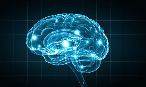
Biopsychology research methods study the human brain. Thanks to them, it’s now easier to understand how the most mysterious organ in our body works. But what exactly are these methods?
Biopsychology Research Methods

Biopsychology research methods have evolved in the last decades. Although there are several biopsychology research methods, in this article, we”ll focus on those that study what happens in the brain under certain conditions.

Authors such as Dewsbury (1991) define biopsychology as “the scientific study of the biology of behavior”, a field also called psychobiology. However, other authors prefer the term biopsychology because it “indicates a biological approach to the study of psychology, more than a psychological approach to the study of biology.”
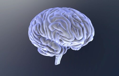
The brain and biopsychology research methods.

Human brain stimulation and visualization methods

Observing and recording brain activity is a very important milestone that was achieved thanks to the different techniques that scientists developed during the 20th century. These biopsychology research methods are, without a doubt, a breakthrough in the study of our most curious organ.

Contrast radiography

This technique consists of injecting a substance in the body to absorb X-rays. This way, scientists see the contrast between the compartment and the tissue around it.

Cerebral angiography is a kind of contrast radiography. To do it, a contrast medium is inserted into a brain vessel. The aim is to observe the circulatory system while performing an X-ray. This technique is very useful to locate vessel injuries and brain tumors.

Computerized axial tomography scan (CT scan)

Through a CT scan, experts can see the whole brain structure. During the test, the patient lies down in the middle of a big cylinder. While the patient holds still, an X-ray tube and a receptor take many separate photographs. This happens while the emitter and the receptor spin around the patient”s head.

All this information is transmitted to a computer, which allows doctors to explore the brain on a horizontal plane. Usually, they do it on eight to nine horizontal brain sections. Once all explorations are combined, it”s possible to make a three-dimensional representation of the brain.

Nuclear magnetic resonance (NMR)

NMR facilitates high-resolution imaging thanks to the different waves hydrogen atoms emit when activated by radio frequency on a magnetic field. It provides a high spatial resolution and produces three-dimensional images.

Positron emission tomography (PET)

PET provides brain activity images instead of brain structure images. To get the images, scientists inject radioactive fludeoxyglucose (FDG) in the carotid artery. Active neurons rapidly absorb FDG, which accumulates once neurons don”t metabolize it anymore, then it slowly degrades. This is how one can observe which neurons are active at a given time during different activities.

Functional magnetic resonance imaging (fMRI)

On the other hand, MRIs offer an image of the increase in the amount of oxygen present in the brain”s blood. Thus, it successfully measures brain activity. If we compare it to the PET, it actually has four advantages:

  • Doctors don”t have to inject the patient with anything.
  • It provides both functional and structural information.
  • It offers a better spatial resolution.
  • Provides three-dimensional images of the entire brain.
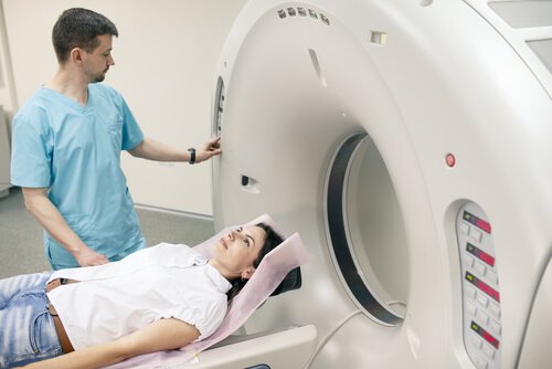
Woman doing MRI test.

Magnetoencephalography (MEG)

It measures the changes in the magnetic fields located on the scalp”s surface. These changes occur due to the variations in the neuronal activity guidelines.

Transcranial magnetic stimulation (TMS)

Walsh and Rothwell (2000) state that TMS “alters an area of the cortex, creating a magnetic field under the coil that goes over the cranium.” TMS basically “turns off” a part of the brain temporarily, to study behavior and cognition under those circumstances.

Lesion methods

Lesion methods focus on the destruction of a small brain area to see how it affects behavior.

  • Aspiration lesion. This method creates a lesion in an exposed or easily accessible cortical tissue area. The doctors remove the tissue with a fine-point crystal pipette.
  • Radio-frequency lesion. It”s carried out by creating small subcortical lesions. For that, an electrode channels the high-frequency current through the tissue of interest. The size and shape of the lesion depend on three factors:
    • Duration of the procedure.
    • Intensity of the current.
    • Configuration of the electrode”s point.
  • Scalpel cuts. It consists of sectioning the brain area of interest.
  • Cooling lesion. This biopsychology research method, although included under lesion methods, is actually temporary and reversible. Instead of destroying tissue, one area is cooled a bit over the freezing point. Neurons stop emitting signals, so the cold brain area remains blocked. With this, researchers are able to see what behavior alterations are caused by those areas. Once the temperature goes back to normal, brain function is restored.

Electrical stimulation

Another biopsychology research method is electrical stimulation. The procedure consists of electrically stimulating a nervous system structure to get data about its functions. Usually, a bipolar electrode is used.

This stimulation “shoots” neurons and alters their behavior. In general, it tends to get the opposite effect of lesion methods. For example, if it”s possible to drastically reduce sleep hours with a lesion, then sleep behavior can become inconvenient and harder to control with electrical stimulation.

Lesion methods with electrical recording

  • Intracellular recording. This technique is carried out by introducing a microelectrode in the interior of a neuron. It records fluctuations of membrane potential.
  • Extracellular unit recording. A microelectrode is placed in the extracellular fluid that surrounds the neuron. It doesn”t provide information on membrane potential.
  • Multi-unit recording. In this case, the electrode point is bigger than that of a microelectrode, so it captures the signals of many neurons at the same time. The detected potentials then go on a circuit that integrates them.
  • Invasive EEG monitoring. The stainless steel electrodes go inside the cranium. For subcortical signals, the usual electrodes are made of cable and implanted through stereotactic surgery.

“Anthropology, biology, physiology and psychology have accumulated mountains of material to raise up before mankind in their full scope the tasks of perfecting and developing body and spirit.”

-Leon Trotsky-
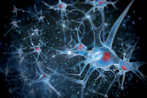
Neurons.

Biopsychology research methods: A long way to go

In this article, we talked about the most important biopsychology research methods. However, it”s also worth mentioning that there are other biopsychology research methods that study other body areas, such as muscle tension measurements, eye movement recordings, skin conductance, or cardiovascular activity.

Without a doubt, the breakthrough in this field has been spectacular, but not conclusive. Perhaps in a few years, scientists will come up with new techniques that will contribute to the evolution of neuroscience, which will help improve the quality of life of many people affected by neuronal alterations.

The Six Major Divisions of Biopsychology

  1. Physiological Psychology – Study of the neural mechanisms of behaviour by manipulating the nervous  systems of nonhuman  animals in controlled experiments 
  2. Psychopharmacology – Study of the effects of drugs on the brain and behaviour
  3. Neuropsychology – Study of the psychological effects of brain damage in human  patients 
  4. Psychophysiology – Study of the relation between physiological activity and psychological process in  human  subjects by noninvasive physiological recording o Noninvasive  – physiological activity is recorded from the surface  of the  body (i.e. EEG – from the scalp)
  5. Cognitive Neuroscience – Study of neural mechanisms in human cognition, largely through the use of  functional brain imaging o Functional brain imaging  – recording the activity of the brain by taking  images of the brain while a subject is performing a particular cognitive  activity
  6. Comparative Psychology – Studies the biology of behaviour rather than the neural mechanisms of behaviour – Compare the behaviour of different species in order to understand the evolution,  genetics, and adaptiveness of behaviour
  • converging Operations: How Do Biopsychologists Work Together? – Converging operations – When different approaches come together to focus on a single problem so that the most progress is made o The strengths of one approach compensates for the weaknesses of the others.
  • Scientific Inference: How Do Biopsychologists Study the Unobservable Workings of the Brain? – The empirical (observational) methods that biopsychologists and scientists use to study the unobservable is called scientific inference.

REQUEST AN APPOINTMENT OR BOOK A CONSULANT – Sargam.dange.18@gmail.com

Perception

REQUEST AN APPOINTMENT OR BOOK A CONSULANT – Sargam.dange.18@gmail.com

  • Perceptual processing,
  • Role of attention in perception,
  • Perceptual organization,
  • Perceptual sets,
  • Perceptual constancies,
  • Depth perception,
  • Illusions.

Perception is the sensory experience of the world. It involves both recognizing environmental stimuli and actions in response to these stimuli.

Through the perceptual process, we gain information about the properties and elements of the environment that are critical to our survival. Perception not only creates our experience of the world around us; it allows us to act within our environment.

What Is Perception?

Perception includes the five senses; touch, sight, sound, smell, and taste. It also includes what is known as proprioception, a set of senses involving the ability to detect changes in body positions and movements. It also involves the cognitive processes required to process information, such as recognizing the face of a friend or detecting a familiar scent.

Learn more about how we go from detecting stimuli in the environment to actually taking action based on that information.

Types of Perception

Some of the main types of perception include:

  • Vision 
  • Touch
  • Sound
  • Taste
  • Smell

There are also other senses that allow us to perceive things such as balance, time, body position, acceleration, and the perception of internal states. Many of these are multimodal and involve more than one sensory modality. Social perception, or the ability to identify and use social cues about people and relationships, is another important type of perception.

How It Works

The perceptual process is a sequence of steps that begins with the environment and leads to our perception of a stimulus and action in response to the stimulus. It occurs continuously, but you do not spend a great deal of time thinking about the actual process that occurs when you perceive the many stimuli that surround you at any given moment.

For example, the process of transforming the light that falls on your retinas into an actual visual image happens unconsciously and automatically. The subtle changes in pressure against your skin that allow you to feel objects occur without a single thought.

Perception acts as a filter that allows us to exist and interpret the world without becoming overwhelmed by the abundance of stimuli.

Steps in the Perceptual Process

  1. The Environmental Stimulus
  2. The Attended Stimulus
  3. The Image on the Retina
  4. Transduction
  5. Neural Processing
  6. Perception
  7. Recognition
  8. Action

Impact of Perception

In order to see the impact of perception, it can be helpful to look at how the process works. This varies somewhat for every sense. In the case of visual perception:

  1. The environmental stimulus: The world is full of stimuli that can attract attention through various senses. The environmental stimulus is everything in the environment that has the potential to be perceived.
  2. The attended stimulus: The attended stimulus is the specific object in the environment on which attention is focused.
  3. The image on the retina: This involves light actually passing through the cornea and pupil and onto the lens of the eye. The cornea helps focus the light as it enters the eye, and the iris of the eye controls the size of the pupils in order to determine how much light to let in. The cornea and lens act together to project an inverted image onto the retina.
  4. Transduction: The image on the retina is then transformed into electrical signals in a process known as transduction. This allows the visual messages to be transmitted to the brain to be interpreted.
  5. Neural processing: The electrical signals then undergo neural processing. The path followed by a particular signal depends on what type of signal it is (i.e. an auditory signal or a visual signal).
  6. Perception: In this step of the process, you perceive the stimulus object in the environment. It is at this point that you become consciously aware of the stimulus.
  7. Recognition: Perception doesn’t just involve becoming consciously aware of the stimuli. It is also necessary for the brain to categorize and interpret what you are sensing. The ability to interpret and give meaning to the object is the next step, known as recognition.
  8. Action: The action phase of perception involves some type of motor activity that occurs in response to the perceived and recognized stimulus. This might involve a major action, like running toward a person in distress, or something as subtle as blinking your eyes in response to a puff of dust blowing through the air.

The perceptual process allows you to experience the world around you and interact with it in ways that are both appropriate and meaningful.

Take a moment to think of all the things you perceive on a daily basis. At any given moment, you might see familiar objects in your environment, feel the touch of objects and people against your skin, smell the aroma of a home-cooked meal, and hear the sound of music playing in your next-door neighbor’s apartment. All of these things help make up your conscious experience and allow you to interact with the people and objects around you.

Tips and Tricks

There are some things that you can do that might help you perceive more in the world around you—or at least focus on the things that are important.

  • Pay attention. Perception requires you to attend to the world around you. This might include anything that can be seen, touched, tasted, smelled, or heard. It might also involve the sense of proprioception, such as the movements of the arms and legs or the change in position of the body in relation to objects in the environment.
  • Make meaning of what you perceive. The recognition stage is an essential part of perception since it allows you to make sense of the world around you. By placing objects in meaningful categories, you are able to understand and react appropriately.
  • Take action. The final step of the perceptual process involves some sort of action in response to the environmental stimulus. This could involve a variety of actions, such as turning your head for a closer look or turning away to look at something else.

Potential Pitfalls

The perceptual process does not always go smoothly and there are a number of things that may interfere with perception. Perceptual disorders are cognitive conditions that are marked by an impaired ability to perceive objects or concepts.

Some disorders that may affect perception include:

  • Spatial neglect syndromes, which involve not attending to stimuli on one side of the body
  • Prosopagnosia, a disorder that makes it difficult to recognize faces
  • Aphantasia, a condition characterized by an inability to visualize things in your mind
  • Schizophrenia, which is marked by abnormal perceptions of reality2

Some of these conditions may be influenced by genetics while others result from stroke or brain injury.

History of Perception

Interest in perception dates back to the time of the ancient Greek philosophers who were interested in how people know the world and gain understanding.

As psychology emerged as a science separate from philosophy, researchers became interested in understanding how different aspects of perception worked, particularly the perception of color. In addition to understanding the basic physiological processes that occur, psychologists were also interested in understanding how the mind interprets and organizes these perceptions. The Gestalt psychologists proposed a holistic approach, suggesting that the sum equals more than the sum of its parts. 

Cognitive psychologists have also worked to understand how motivations and expectations can play a role in the process of perception.

Today, researchers also work to investigate perception on the neural level and look at how injury, conditions, and substances might affect perception.

The Perceptual Process

Perception is how you interpret the world around you and make sense of it in your brain. You do so via stimuli that affect your different senses — sight, hearing, touch, smell, and taste. How you combine these senses also makes a difference. For example, in one study, consumers were blindfolded and asked to drink a new brand of clear beer. Most of them said the product tasted like regular beer. However, when the blindfolds came off and they drank the beer, many of them described it as “watery” tasting (Ries, 2009). This suggests that consumers’ visual interpretation alone can influence their overall attitude towards a product or brand.

Graphic depicting sensory receptors processing environmental stimuli and further processing stimuli through Exposure, Attention, Interpretation, and eventually Adaptation.
The perceptual process begins when our sensory receptors (eyes, ears, tongue, nose, and skin) come in contact with sensory stimuli (sights, sounds, tastes, odours, and textiles) around us. Through our sensory system, we are exposed to an infinite amount of stimuli, some of which we pay attention to, and some we tune out completely. Those that receive our attention we evaluate and interpret their meaning based on our methods of cognitive and behavioural processing. After time, some of these stimuli become adapted and we seize to remark on their significance.

REQUEST AN APPOINTMENT OR BOOK A CONSULANT – Sargam.dange.18@gmail.com

On the role of selective attention in visual perception

What is the role of selective attention in visual perception? Before answering this question, it is necessary to differentiate between attentional mechanisms that influence the identification of a stimulus from those that operate after perception is complete. Cognitive neuroscience techniques are particularly well suited to making this distinction because they allow different attentional mechanisms to be isolated in terms of timing and/or neuroanatomy. The present article describes the use of these techniques in differentiating between perceptual and postperceptual attentional mechanisms and then proposes a specific role of attention in visual perception. Specifically, attention is proposed to resolve ambiguities in neural coding that arise when multiple objects are processed simultaneously. Evidence for this hypothesis is provided by two experiments showing that attention—as measured electrophysiologically—is allocated to visual search targets only under conditions that would be expected to lead to ambiguous neural coding.

Gestalt Laws of Perceptual Organization

eveloped by German psychologists, the Gestalt laws describe how we interpret the complex world around us. They explain why a series of flashing lights appear to be moving. And why we read a sentence like this, notli ket his ort hat. These are just a few real-lie examples of the Gestalt laws.

gestalt laws of perceptual organization
Verywell / JR Bee

History of the Gestalt Laws

Have you noticed how alternately flashing lights, such as neon signs or strands of lights, can look like a single light that was moving back and forth? This optical illusion is known as the phi phenomenon. Discovered by German psychologist Max Wertheimer, this illusion of movement became a basis for Gestalt psychology.

According to Gestalt psychology, this apparent movement happens because our minds fill in missing information. Motion pictures are based on this principle, with a series of still images appearing in rapid succession to form a seamless visual experience.

Gestalt psychology focuses on how our minds organize and interpret visual data.2 It emphasizes that the whole of anything is greater than its parts.

Based upon this belief, Wertheimer along with Gestalt psychologists Wolfgang Köhler and Kurt Koffka, developed a set of rules to explain how we group smaller objects to form larger ones (perceptual organization). They called these rules the Gestalt Laws.

It’s important to note that while Gestalt psychologists call these phenomena “laws,” a more accurate term would be “principles.” These principles are much like heuristics, which are mental shortcuts for solving problems.

Law of Similarity

The law of similarity states that similar things tend to appear grouped together. Grouping can occur in both visual and auditory stimuli.

In the image at the top of this page, for example, you probably see two separate groupings of colored circles as rows rather than just a collection of dots.

Law of Prägnanz

The law of prägnanz is sometimes referred to as the law of good figure or the law of simplicity. This law holds that when you’re presented with a set of ambiguous or complex objects, your brain will make them appear as simple as possible.3 For example, when presented with the Olympic logo, you see overlapping circles rather than an assortment of curved, connected lines.

The word prägnanz is a German term meaning “good figure.”

Law of Proximity

According to the law of proximity, things that are close together seem more related than things that are spaced farther apart.4

In the image at the top of the page, the circles on the left appear to be part of one grouping while those on the right appear to be part of another. Because the objects are close to each other, we group them together.

Law of Continuity

The law of continuity holds that points that are connected by straight or curving lines are seen in a way that follows the smoothest path. In other words, elements in a line or curve seem more related to one another than those positioned randomly.

Law of Closure

According to the law of closure, we perceive elements as belonging to the same group if they seem to complete some entity.1 Our brains often ignore contradictory information and fill in gaps in information.

In the image at the top of the page, you probably see the shape of a diamond because your brain fills in the missing gaps in order to create a meaningful image.

The Law of Common Region

The Gestalt law of common region says that when elements are located in the same closed region, we perceive them as belonging to the same group.1

Look at the last image at the top of the page. The circles are right next to each other so that the dot at the end of one circle is actually closer to the dot at the end of the neighboring circle. But despite how close those two dots are, we see the dots inside the circles as belonging together.

Creating a clearly defined boundary can overpower other Gestalt laws such as the law of proximity.

A Word From Verywell

The Gestalt laws of perceptual organization present a set of principles for understanding some of the ways in which perception works. Research continues to offer insights into perception and how we see the world. These principles of organization play a role in perception, but it is also important to remember that they can sometimes lead to incorrect perceptions of the world.

It is important to remember that while these principles are referred to as laws of perceptual organization, they are actually heuristics or shortcuts. Heuristics are usually designed for speed, which is why our perceptual systems sometimes make mistakes and we experience perceptual inaccuracies.

Perceptual Sets in Psychology

A perceptual set refers to a predisposition to perceive things in a certain way. In other words, we often tend to notice only certain aspects of an object or situation while ignoring other details.

What Is a Perceptual Set?

When it comes to our perceptions of the world around us, you might assume that what you see is what you get. However, in truth, research shows that the way you see the world is heavily influenced (and biased) by your own past experiences, expectations, motivations, beliefs, emotions, and even your culture.1

For example, think about the last time you started a new class. Did you have any expectations at the outset that might have influenced your experience in the class? If you expect a class to be boring, are you more likely to be uninterested in class?

In psychology, this is what is known as a perceptual set.

A perceptual set is basically a tendency to view things only in a certain way.

Perceptual sets can impact how we interpret and respond to the world around us and can be influenced by a number of different factors.

What exactly is a perceptual set, why does it happen, and how does it influence how we perceive the world around us?

How It Works

How do psychologists define perceptual sets?

“Perception can also be influenced by an individual’s expectations, motives, and interests. The term perceptual set refers to the tendency to perceive objects or situations from a particular frame of reference,” explains author Sandra Hockenbury the textbook Discovering Psychology.

Sometimes, perceptual sets can be helpful. They often lead us to make fairly accurate conclusions about what exists in the world around us. In cases where we find ourselves wrong, we often develop new perceptual sets that are more accurate.

Sometimes, our perceptual sets can lead us astray.

If you have a strong interest in military aircraft, for example, an odd cloud formation in the distance might be interpreted as a fleet of fighter jets.

In one experiment that illustrates this tendency, participants were presented with different non-words, such as sael. Those who were told that they would be reading boating-related words read the word as “sail,” while those who were told to expect animal-related words read it as “seal.”

Top-Down Processing

A perceptual set is a good example of what is known as top-down processing. In top-down processing, perceptions begin with the most general and move toward the more specific. Such perceptions are heavily influenced by expectations and prior knowledge.5

If we expect something to appear in a certain way, we are more likely to perceive it according to our expectations.

Existing schemas, mental frameworks, and concepts often guide perceptual sets. For example, people have a strong schema for faces, making it easier to recognize familiar human faces in the world around us. It also means that when we look at an ambiguous image, we are more likely to see it as a face than some other type of object.

Researchers have also found that when multiple items appear in a single visual scene, perceptual sets will often lead people to miss additional items after locating the first one. For example, airport security officers might be likely to spot a water bottle in a bag but then miss that the bag also contains a firearm.

Forces of Influence 

Below are examples of various forces of influence:

  • Motivation can play an important role in perceptual sets and how we interpret the world around us. If we are rooting for our favorite sports team, we might be motivated to view members of the opposing team as overly aggressive, weak, or incompetent. In one classic experiment, researchers deprived participants of food for several hours. When they were later shown a set of ambiguous images, those who had been food-deprived were far more likely to interpret the images as food-related objects.6 Because they were hungry, they were more motivated to see the images in a certain way.
  • Expectations also play an important role. If we expect people to behave in certain ways in certain situations, these expectations can influence how we perceive these people and their roles. One of the classic experiments on the impact of expectation on perceptual sets involved showing participants either a series of numbers or letters. Then, the participants were shown an ambiguous image that could either be interpreted as the number 13 or the letter B. Those who had viewed the numbers were more likely to see it as a 13, while those who had viewed the letters were more likely to see it as the letter B.
  • Culture also influences how we perceive people, objects, and situations. Surprisingly, researchers have found that people from different cultures even tend to perceive perspective and depth cues differently.
  • Emotions can have a dramatic impact on how we perceive the world around us. For example, if we are angry, we might be more likely to perceive hostility in others. One experiment demonstrated that when people came to associate a nonsense syllable with mild electrical shocks, they experienced physiological reactions to the syllable even when it was presented subliminally.
  • Attitudes can also have a powerful influence on perception. In one experiment, Gordon Allport demonstrated that prejudice could have an influence on how quickly people categorize people of various races.

Real-Life Examples

Researchers have shown that perceptual sets can have a dramatic impact on day-to-day life.

In one experiment, young children were found to enjoy French fries more when they were served in a McDonald’s bag rather than just a plain white bag.11 In another study, people who were told that an image was of the famed “Loch Ness monster” were more likely to see the mythical creature in the image, while others who later viewed the image saw only a curved tree trunk.

As previously mentioned, our perceptual set for faces is so strong that it actually causes us to see faces where there are none. Consider how people often describe seeing a face on the moon or in many of the inanimate objects that we encounter in our everyday lives.

REQUEST AN APPOINTMENT OR BOOK A CONSULANT – Sargam.dange.18@gmail.com

Perceptual Constancy Defined

Perceptual constancy refers to the tendency to perceive an object you are familiar with as having a constant shape, size, and brightness despite the stimuli changes that occur. But what does this really mean?

Suppose you are at a neighborhood bus stop with a friend. You see the bus as it turns the corner a few blocks away. From a distance, the bus looks like a mere dot in your field of vision. You put up your palm and notice that you can cover the entire area of the bus with your palm. As the bus approaches the stop, it begins to take up more and more of your field of vision. Your palm no longer covers the area of the bus.

By the time the bus reaches the stop, you realize that the bus is twice as tall as you. Despite the fact that the bus now takes up a majority of your field of vision, you don’t perceive the bus as having grown. You know that the bus has the same size, rectangular shape, and brightness now as it did when you saw it in the distance. The reason you know this is due to perceptual constancy.

Even though the image of the bus gets bigger in size, you have enough experiences with buses to know that the actual size of the bus remains constant. You are able to perceive the size of the bus as stable even though the features of the bus continually change in your field of vision. It is perceptual constancy that allows you to identify objects under different conditions, as you mentally reconstruct images of these objects. For example, the bus appears to be the same size in sunlight during the summer, as well as during a dark, snowy, evening in the winter.

Perceptual constancy can be reduced if you are not very familiar with the object, or if there is a decrease in the number of environmental cues to assist with the object’s identification. So what are the different types of perceptual constancies?

Everything to Know About Depth Perception Issues

When people talk about depth perception, they’re referring to your eyes’ ability to judge the distance between two objects.

Both of your eyes perceive the same object slightly differently and at slightly different angles, but your brain can merge the two images into one 3-D image. This process is also known as stereopsis.

With this information, you can gauge how far apart the objects are, as well as how far they are from you.

How to test depth perception

You can test your eyes’ ability to provide this information at home. Here’s how to do it:

  1. Gaze at a picture of a circle or a ball.
  2. Then, hold up one finger about 6 inches away from your eyes, with the circle in the background.
  3. Focus both eyes on your finger. You’ll probably notice slightly hazy images of the circle that appear on both sides of your finger.
  4. Now, switch your focus. Keep your finger in the same place, but gaze beyond your finger to the circle.
  5. You should see images of your finger on either side of the circle.

If you’re having trouble, you may have an issue with your depth perception.

Causes of depth perception issues

A number of factors can contribute to problems with depth perception. Some of the most common factors include:

Strabismus

Strabismus is a condition that occurs when your eyes are not properly aligned. For example, one eye might turn inward or outward. Sometimes, an eye can also look upward or downward. Some people appear to have crossed eyes, which is also a sign of strabismus.

Essentially, since your eyes may look in slightly different directions, they’ll focus on different things.

The American Academy of Ophthalmology (AAO) estimates that strabismus affects about 4 percent of children in the United States.

Ambylopia

If your brain favors one eye over the other, resulting in one eye that doesn’t quite track properly, you have amblyopia.

Also known colloquially as “lazy eye,” amblyopia can cause vision loss in the weaker eye that can reduce your depth perception and maybe even your vision. It’s also relatively common in babies and young children, according to the AAO.

Nerve problems

If your optic nerve is swollen or inflamed, it might affect your vision and interfere with your depth perception.

Additionally, some people are born with a rare type of nerve damage called optic nerve hypoplasia, which occurs when the optic nerve doesn’t fully develop.

Trauma to one of your eyes

If one of your eyes has been injured, you may no longer be able to see well enough to have good depth perception.

Blurry vision

There are dozens of potential causes of blurry vision, from corneal abrasions and glaucoma to diabetic retinopathy and plain old nearsightedness.

Any condition that makes your vision blurry, even temporarily, can interfere with your ability to perceive distances and depth accurately.

How it impacts daily life

You use visual cues to make all sorts of decisions every single day. And you probably don’t even think about it unless something is wrong.

When something affects your depth perception, it can also interfere with your daily life.

Here are a few common examples of how a problem that affects your depth perception may create some challenges.

Kids and learning

Children who can’t see very well may not say anything about it. But a careful observer may notice them squinting or moving their head in an effort to try to get a better view.

Some children may even have trouble learning because they can’t see the board or other teaching materials at school.

Adults and driving

People with impaired vision or no vision in one eye may worry about how they’re going to get around. The good news is that you can probably still get a driver’s license.

However, because your vision problems can affect or reduce your depth perception abilities, you may need to use some strategies to help you drive safely.

Navigating the world around you

Depth perception helps professional baseball players gauge the speed of the ball hurtling toward them. But depth perception also helps people perform simple, everyday tasks, like safely crossing a busy street or taking the stairs without the risk of misjudging them and stumbling.

If your depth perception is impaired, you may have some trouble doing those kinds of activities. You might even have trouble pouring a glass of milk.

Treatments

The treatment options for depth perception issues depend on the cause of the problem.

For example, children who have strabismus have trouble with depth perception because their eyes are misaligned. So, glasses can help some of them by helping straighten their eyes.

However, other children may need surgery to straighten their eyes, and they might still need to wear glasses after the surgery.

In other cases, such as when a child has amblyopia, a doctor might recommend putting a patch over the good eye for short periods of time to strengthen the weaker eye.

If you have another type of eye condition that impairs your depth perception, talk to your eye doctor about the best possible treatment options. You might need surgery to remove a cataract, for example, or treatment for glaucoma.

Other tips to cope

Some people with limited sight in one eye but good sight in the other are able to adapt. They get enough visual information from their good eye to be able to perceive depths and make judgments based on what that they do receive.

For example, drivers can monitor factors like the relative speed of other cars on the road and how light affects cars and other objects on or near the roadway. These spatial cues can help you gauge the distance between your car and other vehicles.

There are other strategies to try, too. For example, before driving, find ways to reduce glare and anything else that can interfere with your ability to see as best you can.

This might include cleaning your windshield to eliminate any grime or dirt that can interfere with your view. You can also dim the rearview mirror and angle your side mirrors to minimize the glare from the headlights of cars behind you.

Products that can help

Coping strategies can definitely help, but you might also benefit from the use of certain products or services.

These may include:

Vision therapy

Children can often benefit from vision therapy to help train their brains and their eyes to gain or regain better vision. In some cases, the training may involve physical exercises.

Eye patching can also be a part of vision therapy. Adults with certain eye conditions may also benefit from vision therapy.

Specialized glasses

If you plan to drive, you may be a candidate for specialized glasses that can help you. Your doctor may suggest using bioptic telescope attachments on your glasses to make it easier for you to see things that are farther away.

Better lighting

If moving around your home is a challenge, try altering the lighting and contrast in certain areas to help you see better.

For example, if you have trouble perceiving depth differences at night, try improving the lighting in and around your home, so you’re not creeping around in the dark.

Additionally, placing brightly colored tape on the edge of your stairs may help you navigate them with less fear.

When to talk with your doctor

If you’ve long since adapted to an eye condition that affects your depth perception, you may have already developed a toolbox of effective ways to compensate. But if you’ve just recently sustained an eye injury or developed a condition that’s affecting your vision, it’s a good idea to talk to your eye doctor.

You may not be aware of any depth perception issues yet, but your doctor may be able to examine you and determine if it’s something you should be monitoring.

The bottom line

It’s easy to take good depth perception for granted. But you can still live a full life even if your depth perception has been impaired in some way. You may benefit from certain treatments or vision therapy, or you may find aids to help you.

Many people with depth perception issues develop a combination of strategies to help them go about their lives as best they can. If you notice any changes to your vision, be sure to check with your doctor, just in case you’re experiencing any new problems that may need to be treated.

REQUEST AN APPOINTMENT OR BOOK A CONSULANT – Sargam.dange.18@gmail.com

Carpal tunnel syndrome.

REQUEST AN APPOINTMENT OR BOOK A CONSULANT – Sargam.dange.18@gmail.com

Carpal tunnel syndrome is the compression of the median nerve as it passes into the hand. The median nerve is located on the palm side of your hand (also called the carpal tunnel). The median nerve provides sensation (ability to feel) to your thumb, index finger, long finger, and part of the ring finger. It supplies the impulse to the muscle going to the thumb. Carpal tunnel syndrome can occur in one or both of your hands.

Swelling inside your wrist causes the compression in carpal tunnel syndrome. It can lead to numbness, weakness, and tingling on the side of your hand near the thumb.

Carpal tunnel syndrome is caused by pressure on the median nerve. The carpal tunnel is a narrow passageway surrounded by bones and ligaments on the palm side of your hand. When the median nerve is compressed, the symptoms can include numbness, tingling and weakness in the hand and arm.

The anatomy of your wrist, health problems and possibly repetitive hand motions can contribute to carpal tunnel syndrome.

Proper treatment usually relieves the tingling and numbness and restores wrist and hand function.

causes

your wrist and on the median nerve. Inflammation can cause swelling. The most common cause of this inflammation is an underlying medical condition that causes swelling in the wrist, and sometimes obstructed blood flow. Some of the most frequent conditions linked with carpal tunnel syndrome are:

  • diabetes
  • thyroid dysfunction
  • fluid retention from pregnancy or menopause
  • high blood pressure
  • autoimmune disorders such as rheumatoid arthritis
  • fractures or trauma to the wrist

Carpal tunnel syndrome can be made worse if the wrist is overextended repeatedly. Repeated motion of your wrist contributes to swelling and compression of the median nerve. This may be the result of:

  • positioning of your wrists while using your keyboard or mouse
  • prolonged exposure to vibrations from using hand tools or power tools
  • any repeated movement that overextends your wrist, such as playing the piano or typing

Pathophysiology

There is a wide spectrum of causative pathologies, converging on two mechanisms of disease, both of which lead to entrapment:

  • A decrease in the size of the carpal tunnel caused by such conditions as:
    • Mechanical overuse (considered the most common association)
    • osteoarthritis
    • Trauma
    • acromegaly
  • Disease states leading to augmentation of carpal tunnel contents:
    • Masses, For example, ganglion cysts, primary nerve sheath tumours
    • Deposition of foreign material, e.g. amyloid
    • Synovial hypertrophy in rheumatoid arthritis

In general, the pathophysiology of CTS results from a combination of compression and traction mechanisms. 

  1. The compressive element of the pathophysiology includes a detrimental cycle of increased pressure, obstruction of overall venous outflow, increasing local edema, and compromise to the median nerve’s intraneural microcirculation.  Nerve dysfunction becomes compromised, and the structural integrity of the nerve itself further propagates the dysfunctional environment– the myelin sheath and axon develop lesions, and the surrounding connective tissues become inflamed and lose normal physiologic protective and supportive function. 
  2. Repetitive traction and wrist motion exacerbates the negative environment, further injuring the nerve.  In addition,  any of the nine flexor tendons traveling through the carpal tunnel can become inflamed and compress the median nerve

Idiopathic Carpal Tunnel Syndrome

  • Idiopathic CTS occurs more frequently in females (65–80%), between the ages of 40 and 60 years; 50–60% of the cases are bilateral.
  • The bilateral characteristic increases in frequency with the duration of symptoms. 
  • Idiopathic CTS is correlated with hypertrophy of the synovial membrane of the flexor tendons caused by degeneration of the connective tissue, with vascular sclerosis, edema and collagen fragmentation.
  • The histological changes were thought to be suggestive of dynamic factors as repetitive strain.

Clinically Relevant Anatomy

Fig.2 Transverse plane view of carpal tunnel.

The carpal tunnel (CT) is formed by a non-extendable osteofibrous wall that forms a tunnel protecting the median nerve and flexor tendons.

The transverse carpal ligament (flexor retinaculum) makes up the superior boundary, and the carpal bones form the inferior border.

The carpal tunnel includes the median nerve and nine flexor tendons.

The flexor tendons includeFour tendons from the flexor digitorum profundus

Four tendons from the flexor digitorum superficialis

One tendon from the flexor pollicis longus.

Symptoms

Carpal tunnel syndrome symptoms usually start gradually and include:

  • Tingling or numbness. You may notice tingling and numbness in your fingers or hand. Usually the thumb and index, middle or ring fingers are affected, but not your little finger. You might feel a sensation like an electric shock in these fingers. The sensation may travel from your wrist up your arm. These symptoms often occur while holding a steering wheel, phone or newspaper, or may wake you from sleep. Many people “shake out” their hands to try to relieve their symptoms. The numb feeling may become constant over time.
  • Weakness. You may experience weakness in your hand and drop objects. This may be due to the numbness in your hand or weakness of the thumb’s pinching muscles, which are also controlled by the median nerve.

When to see a doctor

See your doctor if you have signs and symptoms of carpal tunnel syndrome that interfere with your normal activities and sleep patterns. Permanent nerve and muscle damage can occur without treatment.

REQUEST AN APPOINTMENT OR BOOK A CONSULANT – Sargam.dange.18@gmail.com

Risk factors

A number of factors have been associated with carpal tunnel syndrome. Although they may not directly cause carpal tunnel syndrome, they may increase the risk of irritation or damage to the median nerve. These include:

  • Anatomic factors. A wrist fracture or dislocation, or arthritis that deforms the small bones in the wrist, can alter the space within the carpal tunnel and put pressure on the median nerve. People who have smaller carpal tunnels may be more likely to have carpal tunnel syndrome.
  • Sex. Carpal tunnel syndrome is generally more common in women. This may be because the carpal tunnel area is relatively smaller in women than in men. Women who have carpal tunnel syndrome may also have smaller carpal tunnels than women who don’t have the condition.
  • Nerve-damaging conditions. Some chronic illnesses, such as diabetes, increase your risk of nerve damage, including damage to your median nerve.
  • Inflammatory conditions. Rheumatoid arthritis and other conditions that have an inflammatory component can affect the lining around the tendons in your wrist and put pressure on your median nerve.
  • Medications. Some studies have shown a link between carpal tunnel syndrome and the use of anastrozole (Arimidex), a drug used to treat breast cancer.
  • Obesity. Being obese is a risk factor for carpal tunnel syndrome.
  • Body fluid changes. Fluid retention may increase the pressure within your carpal tunnel, irritating the median nerve. This is common during pregnancy and menopause. Carpal tunnel syndrome associated with pregnancy generally gets better on its own after pregnancy.
  • Other medical conditions. Certain conditions, such as menopause, thyroid disorders, kidney failure and lymphedema, may increase your chances of carpal tunnel syndrome.
  • Workplace factors. Working with vibrating tools or on an assembly line that requires prolonged or repetitive flexing of the wrist may create harmful pressure on the median nerve or worsen existing nerve damage, especially if the work is done in a cold environment. However, the scientific evidence is conflicting and these factors haven’t been established work as direct causes of carpal tunnel syndrome. Several studies have evaluated whether there is an association between computer use and carpal tunnel syndrome. Some evidence suggests that it is mouse use, and not the use of a keyboard, that may be the problem. However, there has not been enough quality and consistent evidence to support extensive computer use as a risk factor for carpal tunnel syndrome, although it may cause a different form of hand pain.

diagnosis

Doctors can diagnose carpal tunnel syndrome using a combination of your history, a physical examination, and tests called nerve conduction studies.

A physical examination includes a detailed evaluation of your hand, wrist, shoulder, and neck to check for any other causes of nerve pressure. Your doctor will look at your wrists for signs of tenderness, swelling, and any deformities. They will check sensation to the fingers and strength of the muscles in your hand.

Nerve conduction studies are diagnostic tests that can measure the conduction speed of your nerve impulses. If the nerve impulse is slower than normal as the nerve passes into the hand, you may have carpal tunnel syndrome.

Differential Diagnosis

The process of differential diagnosis should give consideration to all conditions which could potentially cause a dysfunction of the median nerve, or its contributories in brachial plexus, C 5 to 8 nerve roots and central nervous system.

Possible differential diagnoses of carpal tunnel syndrome include:

  • other median nerve entrapment syndromes
  • pronator teres syndrome
  • anterior interosseous nerve syndrome
  • an injury of nerve digitales in the palm.
  • cervicobrachial syndrome

Physical Examination

May include testing for sensory and motor deficits and evidence of thenar wasting. There are several special tests with varying degrees of sensitivities and specificities.

  • The best of these include the carpal compression test. This is done by applying firm pressure directly over the carpal tunnel for 30 seconds. The test is positive when paresthesias, pain, or other symptoms are reproduced.
  • The square sign test is an evaluation to determine the risk of developing carpal tunnel syndrome. The test is positive if the ratio of the thickness of the wrist divided by the width of the wrist is great than 0.7.
  • Another test is a palpatory diagnosis. In this test, the health care provider examines soft tissue over the median nerve for mechanical restriction.
  • The Phalen’s test or ‘reverse prayer’ is performed by having the patient fully flex their wrists by placing dorsal surfaces of both hands for one minute. A positive test is when symptoms (numbness, tingling, pain) are reproduced.
  • The reverse Phalen’s, or ‘prayer test,’ is done by having the patient extend both of their wrists by placing palmar surfaces of both hands together for 1 minute (as if praying). Again a positive test is with reproduction of symptoms.
  • Although a low sensitivity and specificity, the Hoffmann-Tinel sign is another test commonly performed. In this test the healthcare professional taps immediately over the carpal tunnel to stimulate the median nerve. Like the above tests, a positive test is when symptoms are reproduced.

Clinical prediction rules (CPR), specifically for the Carpal Tunnel Syndrome, are a reliable examination method.

Medical Management

If carpal tunnel syndrome is identified early conservative treatment is recommended.

  • Initially, the patient should be instructed in modifying symptom provoking wrist movement, For example, proper hand ergonomics such as placing the keyboard at a proper height and minimizing flexion, extension, abduction, and adduction of the hand when typing. It should be recommended to decrease repetitive activities if possible.[1]
  • Non-surgical treatment comprises oral steroids , corticosteroid injections, NSAID, diuretics vitamin B6  and splinting/hand brace . If conservative treatments are not successful, an oral or local glucocorticoid could be offered. 
  • The definitive treatment for persistent carpal tunnel syndrome is surgical intervention with carpal tunnel release after nerve conduction studies showing significant axonal degeneration. Carpal tunnel release typically is performed by an orthopedic surgeon or hand surgeon. This procedure can be performed either open or endoscopically. Carpal tunnel release is considered a minor surgery in which the transverse carpal ligament or flexor retinaculum is cut, opening more space in the carpal tunnel and decreasing pressure on the median nerve. It does not typically require overnight hospitalization.

Physical Therapy Management

Patients with mild to moderate symptoms can be effectively treated in a primary care environment 

Physical therapists should give advice on modifications of activities and the workplace (ergonomic modifications), task modification, For example, taking sufficient rest and variation of movements.

Often simple obvious alterations to the working practice can be beneficial in controlling milder symptoms of CTS.

Manual therapy techniques include mobilisation of

  • Soft tissue
  • Carpal bone
  • Median nerve

Other modalities include: ultrasound and electromagnetic field therapy and splinting.

Research findings (varies)

  • Physiotherapy modalities (TENS and ultrasound) have little useful effects on hand sensory discomfort.
  • The evidence of the effectiveness of the exercise and mobilization interventions is limited and very low in quality..
  • Evidence about post-operative rehabilitation is also limited. None of them seems to have a prevailing benefit.

Clinical Bottom Line

Patients may benefit from physiotherapy based treatment with goals of CTS symptom reduction and functional gains, provided that:

  • Their symptoms are intermittent and not rapidly worsening or if their CTS etiology is highly suggestive of the possibility of remission as an example of pregnancy-related CTS
  • Patients are informed about the lack of high-quality evidence for the effectiveness and safety of therapeutic modalities used by physiotherapists,

Treatment should be discontinued when shown to be ineffective and appropriate, evidence-based discharge recommendations should be made.

CTS symptoms typically escalate over a longer-term despite conservative treatment. Surgical interventions for appropriate patients have been shown to be safe and more effective than any conservative intervention, Clinicians should be aware that constant tingling or numbness is associated with significant compression of the median nerve. Prolonged duration of such symptoms may lead to irreversible changes in its internal structure, affecting the effectiveness of surgery and leaving individuals with chronic symptoms and muscle atrophy of thenar eminence. Patients should be followed by their general practitioners to discuss surgical treatment options when necessary and desired.

REQUEST AN APPOINTMENT OR BOOK A CONSULANT – Sargam.dange.18@gmail.com

Osteochondritis

REQUEST AN APPOINTMENT OR BOOK A CONSULANT – Sargam.dange.18@gmail.com

Osteochondritis dissecans occurs when a fragment of bone in a joint separates from the rest of the bone because its blood supply is faulty, and there is not enough blood to maintain it. It often affects the knee or the elbow.

Sometimes, the separated fragment stays in place or repairs on its own. However, in the later stages, the bone can splinter and fall into the joint space, resulting in pain and dysfunction. These fragments are sometimes called “joint mice.”

The exact prevalence is unknown, but there may be between 15 and 29 cases in every 100,000 people. It is more common in males, especially those between the ages of 10 and 20 years who are physically active.

However, the incidence is increasing in females.

It usually affects teens and young adults, but it can occur in younger children who are active in sports.

Osteochondritis dissecans (os-tee-o-kon-DRY-tis DIS-uh-kanz) is a joint condition in which bone underneath the cartilage of a joint dies due to lack of blood flow. This bone and cartilage can then break loose, causing pain and possibly hindering joint motion.

Osteochondritis dissecans occurs most often in children and adolescents. It can cause symptoms either after an injury to a joint or after several months of activity, especially high-impact activity such as jumping and running, that affects the joint. The condition occurs most commonly in the knee, but also occurs in elbows, ankles and other joints.

Doctors stage osteochondritis dissecans according to the size of the injury, whether the fragment is partially or completely detached, and whether the fragment stays in place. If the loosened piece of cartilage and bone stays in place, you may have few or no symptoms. For young children whose bones are still developing, the injury might heal by itself.

Surgery might be necessary if the fragment comes loose and gets caught between the moving parts of your joint or if you have persistent pain.

Causes

The exact cause is unknown, but they may include:

Ischemia: a restriction of blood supply starves the bone of essential nutrients. The restricted blood supply is usually caused by some problem with blood vessels, or vascular problems. The bone undergoes avascular necrosis, a deterioration caused by lack of blood supply. Ischemia usually occurs in conjunction with a history of trauma.

Genetic factors: OCD sometimes affects more than one family member. This may indicate an inherited genetic susceptibility.

Repeated stress to the bone or joint: this can significantly increase the risk of developing OCD. Individuals who play competitive sports are more likely to regularly stress their joints.

Other factors may be weak ligaments or meniscal lesions in the knee.

Symptoms

Depending on the joint that’s affected, signs and symptoms of osteochondritis dissecans might include:

  • Pain. This most common symptom of osteochondritis dissecans might be triggered by physical activity — walking up stairs, climbing a hill or playing sports.
  • Swelling and tenderness. The skin around your joint might be swollen and tender.
  • Joint popping or locking. Your joint might pop or stick in one position if a loose fragment gets caught between bones during movement.
  • Joint weakness. You might feel as though your joint is “giving way” or weakening.
  • Decreased range of motion. You might be unable to straighten the affected limb completely.

When to see a doctor

If you have persistent pain or soreness in your knee, elbow or another joint, see your doctor. Other signs and symptoms that should prompt a call or visit to your doctor include joint swelling or an inability to move a joint through its full range of motion.

Risk factors

Osteochondritis dissecans occurs most commonly in children and adolescents between the ages of 10 and 20 who are highly active in sports.

Complications

Osteochondritis dissecans can increase your risk of eventually developing osteoarthritis in that joint.

Diagnosis

A person who experiences the symptoms of OCD in a joint should seek medical advice. An early diagnosis can mean more effective treatment and a lower risk of complications.

The doctor will carry out a physical examination and ask the patient about their medical history, family history and lifestyle, including sporting activities.

There may be some imaging tests, such as an X-ray, CT, MRI scan, or ultrasound. This may show whether there is any necrosis, or tissue death, or loose fragments. A bone scan may also be recommended.

In the early stages, tests will show that the cartilage is thickening. In the later stages, there will be loose fragments.

The early stages are considered stable, and treatment is more likely to be effective at this point.

Conditions, with similar symptoms need to be ruled out. These include inflammatory arthritis, osteoarthritis, bone cysts and septic arthritis.

Differential Diagnosis

  • Meniscus and collateral ligament injuries – physical examination can rule this out.

If there is no certain radiological determination of osteochondritis dissecans, there can also be alternative causes of the same symptoms that should be sought for e.g.:

  • Inflammatory arthritites: a group of conditions which affect your own immune system.
  • Osteoarthritis: degradation of joints
  • Bone cysts: type of cyst in joints
  • Septic arthritis: purulent invasion of the knee which produces arthritis
  • Ideopathic osteonecrosis
  • Chondral seperations
  • Osteochondral fractures

An x-ray, ct scan or MRI scan can be performed to show necrosis of subschondral bone or formation of loose fragments. This can lead to a better diagnosis. 

REQUEST AN APPOINTMENT OR BOOK A CONSULANT – Sargam.dange.18@gmail.com

Examination

  • The knee feels warmer than the non-injured knee.
  • There is an intermittent swelling palpable.
  • Quadriceps muscle atrophy
  • The passive and active extension of the knee is limited .
  • Catching or locking of the knee .
  • Tibial external rotation during gait.
  • Fluid effusion
  • It is possible that both capsular and non-capsular movement restrictions can be found during functional assessment, the severity is dependent on a possible herniation of the knee joint and the degree of joint irritation .
  • The sensitive location of the abandoned section of the osteochondral fracture can be felt, when the knee is in 90° of flexion .
  • Wilson’s Test: The knee is held in 90° to 30° from full extension while rotating the tibia.  The test is positive when internal rotation is painful and external rotation relieves symptoms

Prevention

Adolescents participating in organized sports might benefit from education on the risks to their joints associated with overuse. Learning the proper mechanics and techniques of their sport, using the proper protective gear, and participating in strength training and stability training exercises can help reduce the chance of injury.

Diagnosis

A person who experiences the symptoms of OCD in a joint should seek medical advice. An early diagnosis can mean more effective treatment and a lower risk of complications.

The doctor will carry out a physical examination and ask the patient about their medical history, family history and lifestyle, including sporting activities.

There may be some imaging tests, such as an X-ray, CT, MRI scan, or ultrasound. This may show whether there is any necrosis, or tissue death, or loose fragments. A bone scan may also be recommended.

In the early stages, tests will show that the cartilage is thickening. In the later stages, there will be loose fragments.

The early stages are considered stable, and treatment is more likely to be effective at this point.

Conditions, with similar symptoms need to be ruled out. These include inflammatory arthritis, osteoarthritis, bone cysts and septic arthritis.

Treatment

Conservative measures include changes of activity or rest. This can give the bone time to heal and to prevent future fracture, crater formation, or chondral (cartilage) collapse. Rest and the use of crutches may help.

If the patient has been involved in sports, they may need to stop for a while.

The doctor may immobilize the joint with a medical device, such as a splint or a brace. Crutches may be necessary.

A non-steroidal anti-inflammatory medication (NSAID) can help with pain. A physical therapist may offer guidance with stretching and specific exercises.

Children can normally return to sports after 2 to 4 months. In young children, OCD normally heals with rest, as the bones are still growing.

In older children and adults, the effects can be more severe.

Surgery

Surgery aims to:

  • restore normal bloodflow
  • get the joint to work normally again

It may be recommended if conservative measures have not worked, if a lesion has become detached and is moving around inside the joint, or if the lesion is over 1 centimeter in diameter.

Drilling into the lesion can create pathways for new blood vessels to form in. This allows blood to flow, and encourages the bone to heal.

The surgeon makes a small incision. Using some long, thin instruments, they either remove or reattach the loose fragments of bone. If the cartilage is still attached to the bone, pins or screws can be used to secure it.

Osteochondral autograft transfer (OATS) uses healthy cartilage to replace damaged cartilage on the surface of the joint that receives weight-bearing stress. It is like a cartilage transplant, but the recipient and donor is the same person.

After surgery the patient will undergo a rehabilitation program. After an initial period of immobilization, physical therapy can help regain joint strength and stability.

According to the American Academy of Orthopedic Surgeons (AAOS), the patient will probably need:

  • crutches for about 6 weeks after surgery
  • physical therapy for 2 to 4 months, to recover strength and motion

After 4 to 5 months, a gradual return to sports may be possible.

Minimally invasive arthroscopic surgery is less painful, the recovery time is faster, and the risk of complications is lower.

Medical Management

In minor cases rest can be prescribed. The patient has to stop activities for three to six months and the lesion will heal spontaneously, especially with young adolescents.

Normally, immobilization of the knee for a couple of weeks is sufficient in the treatment of growing children. In case immobilization is insufficient, as would normally be the case for adults, a mobilization procedure must be started up. In this procedure, stretching exercises are performed. The range of motion and strengthening ability of the muscles will be gradually increased in the next 3 to 6 months. In the end, in case the knee is not fully recovered, surgery should be necessary. (Level of evidence: C5, F5)

Stages three and four are always treated surgically. Surgery is also required when the conservative treatment in stages one and two was inadequate. It is recommended to treat surgically when a large part of the femoral condyle has been excavated, because of the risk to develop osteoarthritis.

A variety of surgical methods exist for the management of articular cartilage lesions at the knee, such as OCD. These include the use of arthroscopic lavage or debridement, radio frequency energy, bone drilling, osteochondral autografts or allografts, internal fixation of bone fragments, and autologous chondrocyte implantation . (level of evidence: 3B)

Surgical techniques:

  • In stages one and two the articular cartilage is still intact, through retrograde operation trying to tap into to the affected bone ‘from behind’ and clear it. The advantage of this surgical technique is that the articular cartilage stays intact .
  • Not yet dissected fragment will be fixed by means of an operation Excision of the fragment and removal of loose bodies.
  • Repair of blood supply by drilling arthroscopic through the cartilage and the hearth of osteochondrosis into the healthy bones
  • Stabilization of the fragment through pinning or through screw fixation
  • Osteochondral autograft transplantation (OATS).
  • Osteochondral allograft transplantation.
  • Autologous chondrocyte implantation (ACI).

Physical Therapy Management

In stages one and two the condition is localized in the subchondral bone, the cartilage is still intact and gets its nourishment from synovial fluid. In these two stages conservative therapy can be applied. The goals of conservative therapy are: pain reduction, promote the repair of the cartilage and prevent degeneration of the surface of the knee joint.  There is no standard treatment.

Immobilisation

Adaption of the strain is needed so that the bone can heal. 2 weeks of immobilization and partial support is recommended when having an acute injury. With children whose bones will still grow, the bone defect may heal by resting the joint. Long-term immobilization has to be prevented, because joint motion is necessary for the nutrition and strengthening of the cartilage. Sport activities should be stopped temporally.

Physical Therapy

  • Stretching to improve range of motion
  • Strengthening exercises for the muscles
  • First exercises: closed chain exercises, low impact activities like cycle and swim. Using exercises as straight leg raises and ankle band exercises, strength can be maintained.
  • Coactivation or setting of the quadriceps and hamstring can be performed while in an immobilizer or cast.
  • Using neuromuscular electrical stimulation to the quadriceps and hamstrings for coactivation contractions can further augment the strength maintenance program.
  • Following immobilization, range of motion exercises, as well as progressive quadriceps and hamstring strengthening should be performed.
  • Weight-bearing progression throughout rehabilitation should be to patient tolerance.
  • In facilitating the return to full-weight-bearing status is aquatic therapy very beneficial.
  • To adress any gait deviations that developed during the immobilization and decreased weight-bearing phases of rehabilitation gait training techniques may be used, such as manual facilitation and visual feedback tot the patient via a full length mirror.
  • Additional exercises to restore ankle joint and normal knee proprioception, such as biomechanical ankle platform systems (BAPS board) exercises or unilateral stance, are also beneficial to the athlete planning to return to competition.
  • After this period the sport activities can be partly restart.
  • Next criteria should be managed: the patient is pain free, has a full joint mobility, no swelling, no pressure sensitivity and there’s radiological prove of recovery.

Post-operative Physical Therapy

An operative treatment is indicated if, after a treatment of three to six months and no recovery has occurred, or when the loose fragment is to big. The surgery goals would be to remove loose fragments or to reattach fragments.

Immobilization is not necessary before surgery. Immediately after the intervention the knee get scontinuous passive motion for 48 hours. After this therapy is recommend, including 8 weeks of rehabilitation exercises for limb function and recruitment. Between week 6 and 8 weight-bearing is gradually introduced to full weight bearing.

REQUEST AN APPOINTMENT OR BOOK A CONSULANT – Sargam.dange.18@gmail.com

Dietary Assessment

REQUEST AN APPOINTMENT OR BOOK A CONSULANT – Sargam.dange.18@gmail.com

A patient’s nutritional status can be evaluated by using and integrating information obtained from a past medical history, family history and a social history. It is worthwhile to assess the nutritional status of every patient. Many complaints such as fatigue, headaches, and digestive disturbances can be improved by simple changes in diet. Nutrition also plays a pivotal role in prevention. Nutritional assessment becomes especially important when the patient has certain nutritional “red flags”, such as:

  • Elderly
  • No regular exercise regime and/or sedentary lifestyle
  • Recent loss of 10% or more of usual body weight
  • Alcoholism
  • Taking drugs such as steroids, immunosuppressants
  • Infection, protracted fever, trauma
  • Malabsorption syndromes, draining abscesses, renal dialysis
  • Receiving simple intravenous solutions without oral intake for >10 days

Past Medical History

  • Immunizations, hospitalizations, operations, major injuries, chronic illnesses, and significant acute illnesses
  • Current or recent prescription medications, vitamins and minerals, laxatives, topical medications, OTC medications, and nutritional supplements
  • Potential drug-nutrient interactions, such as those caused by potassium-wasting diuretics
  • Food allergies or lactose intolerance

Family History

  • Family history of cancer, diabetes, heart disease, hypertension, obesity, and osteoporosis
  • Parents, siblings, children, spouse: include ages, current health status, and cause of death if deceased

Social History

  • Occupation, daily exercise pattern, marital and family status
  • Economic status, educational level, residence, emotional response to illness and coping skills
  • Duration and frequency of use of substances, including tobacco, alcohol, illegal drugs, and caffeine

Assessing Your Patient’s Dietary Intake

There are several methods for obtaining a dietary history. Each has its advantages and disadvantages.

24-hour Recall

  • An informal, qualitative method in which you ask the patient to recall all of the foods and beverages that were consumed in the last 24 hours, including the quantities and methods of preparation.
  • An advantage of this method is that dietary information is easily obtained. It is also good during a first encounter with a new patient in which there is no other nutritional data. Patients should be able to recall all that they have consumed in the last 24 hours.
  • A disadvantage of this method is that it is very limited and may not represent an adequate food intake for the patient. Data achieved using this method may not represent the long-term dietary habits of the patient. Estimating food quantities and food ingredients may be difficult especially if the patient ate in restaurants.

Usual Intake/Diet History

  • This method asks the patient to recall a typical daily intake pattern, including amount, frequencies and methods of preparation. This intake history should include all meals, beverages and snacks.
  • Advantages of this method are that it evaluates long-term dietary habits and is quick and easy to do. Based on the information acquired, you may identify patients who would benefit from meeting with a registered dietitian.
  • A disadvantage of this method is that a limited amount of information on the actual quantities of food and beverages is obtained. Also, this method only works if a patient can actually describe a “typical” daily intake, which is difficult for those who vary their food intake greatly. In these patients it is would be advisable to use the 24-hour recall method. Another disadvantage is that patients may not include foods that they know are unhealthy.
  • An excellent method to better understand a patient’s nutritional status is to use a usual intake and lifestyle recall. This consists of asking the patient to run through a typical day in chronological order, describing all food consumption as well as activities. This method is very helpful because it may reveal other factors that can affect the patient’s nutritional and overall health. [For example: (1) the patient sits in traffic for a 3-hour round trip each day; (2) the patient comes home from work late and eat dinner just before bedtime; or (3) the most exercise the patient gets is walking to and from the parking lot at work.]

REQUEST AN APPOINTMENT OR BOOK A CONSULANT – Sargam.dange.18@gmail.com

Food Frequency Questionnaire

  • This method makes use of a standardized written checklist where patients check off the particular foods or type of foods they consume. It is used to determine trends in patients’ consumption of certain foods. The checklist puts together foods with similar nutrient content, and frequencies are listed to identify daily, weekly, or monthly consumption.
  • An advantage of this method is that it makes it possible to identify inadequate intake of any food group, so that dietary and nutrient deficiencies may be identified. The questionnaire can be geared to a patient’s pre-existing medical conditions.
  • Disadvantages include patient error in filling out the questionnaire and no way to find out how foods are prepared. Patients may over- or under-estimate food quantities.

Dietary Food Log

  • This method asks the patient to record all food, beverage and snack consumption for a one- week period. Specific foods and quantities should be recorded. The data from the food log may later be entered into a computer program, which will analyze the nutrient components of the foods eaten according to specific name brands or food types. Patients are asked to enter data into the food log immediately after food is consumed so they do not forget.
  • The most important advantage of this method is that a computer can objectively analyze data obtained. Data on calorie, fat, protein and carbohydrate consumption can be obtained. Also, since patients are asked to enter data immediately after eating, the data is more accurate than other methods.
  • Disadvantages include patient error in entering accurate food quantities. In addition, it is possible that the week long food log does not accurately represent a patient’s normal eating habits since they know the foods they eat will be analyzed and therefore may eat healthier.

Generic Nutrition Questionnaire

Do not be daunted by the following list. Reading through the sample questions will simply give you an idea of probing questions you might use to ascertain your patient’s nutritional status.

  1. Would you describe you appetite as hearty, moderate, or poor?
  2. Are you on a special diet? Specify the type of diet. Who recommended the diet? If you have been on a special diet in the past, define it.
  3. Have you ever had any problems with weight? Underweight or overweight?
  4. Do you eat at approximately the same time every day?
  5. Do you skip meals? If so, when?
  6. Do you usually eat between meals? What do you snack on most often?
  7. Are there any foods you do not eat because you don’t think they are good for you? If yes, what?
  8. Are there any foods you do eat regularly because you think they are good for you? If yes, what?
  9. Are there any foods you cannot eat? What happens when you eat this food?
  10. Are there foods you avoid because you don’t like them?
  11. How is your food usually prepared, e.g. baked, broiled, fried?
  12. Are you on a diet now to lose or gain weight? If yes, what kind? Who recommended it?
  13. How do you feel about your weight?
  14. Are you taking any vitamin or mineral supplements? What and how often?
  15. Do you smoke? How many cigarettes (cigars or chewing tobacco) per day?
  16. Do you drink any alcohol? How much per day and when?
  17. How often do you exercise? What types of exercise do you do?
  18. Who in the house plans the meals? Buys the food? Prepares the food?
  19. Are there times in the month when there isn’t enough money for food or you run out of food?
  20. How many of your meals are eaten at home? School? Work? Restaurants?
  21. How many hot meals do you eat per week?
  22. What condiments do you use? Examples include butter on bread, sugar on cereal, salad dressing, gravy, etc.

REQUEST AN APPOINTMENT OR BOOK A CONSULANT – Sargam.dange.18@gmail.com

Gout- Nature and occurrence of uric acid, causes, symptoms and diet

REQUEST AN APPOINTMENT OR BOOK A CONSULANT – Sargam.dange.18@gmail.com

Gout is a painful form of arthritis that occurs when high levels of uric acid in the blood cause crystals to form and accumulate in and around a joint.

Uric acid is produced when the body breaks down a chemical called purine. Purine occurs naturally in your body, but it’s also found in certain foods. Uric acid is eliminated from the body in urine.

A gout diet may help decrease uric acid levels in the blood. A gout diet isn’t a cure. But it may lower the risk of recurring gout attacks and slow the progression of joint damage.

People with gout who follow a gout diet generally still need medication to manage pain and to lower levels of uric acid. click here to read more

Gout diet goals

A gout diet is designed to help you:

  • Achieve a healthy weight and good eating habits
  • Avoid some, but not all, foods with purines
  • Include some foods that can control uric acid levels

A good rule of thumb is to eat moderate portions of healthy foods.

Diet details

The general principles of a gout diet follow typical healthy-diet recommendations:

  • Weight loss. Being overweight increases the risk of developing gout, and losing weight lowers the risk of gout. Research suggests that reducing the number of calories and losing weight — even without a purine-restricted diet — lower uric acid levels and reduce the number of gout attacks. Losing weight also lessens the overall stress on joints.
  • Complex carbs. Eat more fruits, vegetables and whole grains, which provide complex carbohydrates. Avoid foods and beverages with high-fructose corn syrup, and limit consumption of naturally sweet fruit juices.
  • Water. Stay well-hydrated by drinking water.
  • Fats. Cut back on saturated fats from red meat, fatty poultry and high-fat dairy products.
  • Proteins. Focus on lean meat and poultry, low-fat dairy and lentils as sources of protein.

Recommendations for specific foods or supplements include:

  • Organ and glandular meats. Avoid meats such as liver, kidney and sweetbreads, which have high purine levels and contribute to high blood levels of uric acid.
  • Red meat. Limit serving sizes of beef, lamb and pork.
  • Seafood. Some types of seafood — such as anchovies, shellfish, sardines and tuna — are higher in purines than are other types. But the overall health benefits of eating fish may outweigh the risks for people with gout. Moderate portions of fish can be part of a gout diet.
  • High-purine vegetables. Studies have shown that vegetables high in purines, such as asparagus and spinach, don’t increase the risk of gout or recurring gout attacks.
  • Alcohol. Beer and distilled liquors are associated with an increased risk of gout and recurring attacks. Moderate consumption of wine doesn’t appear to increase the risk of gout attacks. Avoid alcohol during gout attacks, and limit alcohol, especially beer, between attacks.
  • Sugary foods and beverages. Limit or avoid sugar-sweetened foods such as sweetened cereals, bakery goods and candies. Limit consumption of naturally sweet fruit juices.
  • Vitamin C. Vitamin C may help lower uric acid levels. Talk to your doctor about whether a 500-milligram vitamin C supplement fits into your diet and medication plan.
  • Coffee. Some research suggests that drinking coffee in moderation, especially regular caffeinated coffee, may be associated with a reduced risk of gout. Drinking coffee may not be appropriate if you have other medical conditions. Talk to your doctor about how much coffee is right for you.
  • Cherries. There is some evidence that eating cherries is associated with a reduced risk of gout attacks.

Sample menu

Here’s what you might eat during a typical day on a gout diet.

Breakfast

  • Whole-grain, unsweetened cereal with skim or low-fat milk
  • 1 cup fresh strawberries
  • Coffee
  • Water

Lunch

  • Roasted chicken breast slices (2 ounces) on a whole-grain roll with mustard
  • Mixed green salad with vegetables, 1 tablespoon nuts, and balsamic vinegar and olive oil dressing
  • Skim or low-fat milk or water

Afternoon snack

  • 1 cup fresh cherries
  • Water

Dinner

  • Roasted salmon (3 to 4 ounces)
  • Roasted or steamed green beans
  • 1/2 to 1 cup whole-grain pasta with olive oil and lemon pepper
  • Water
  • Low-fat yogurt
  • 1 cup fresh melon
  • Caffeine-free beverage, such as herbal tea

Results

Following a gout diet can help limit uric acid production and increase its elimination. A gout diet isn’t likely to lower the uric acid concentration in your blood enough to treat your gout without medication. But it may help decrease the number of attacks and limit their severity.

Following a gout diet, along with limiting calories and getting regular exercise, can also improve your overall health by helping you achieve and maintain a healthy weight.

How Does Food Affect Gout?

If you have gout, certain foods may trigger an attack by raising your uric acid levels.

Trigger foods are commonly high in purines, a substance found naturally in foods. When you digest purines, your body makes uric acid as a waste product.

This is not a concern for healthy people, as they efficiently remove excess uric acid from the body.

However, people with gout can’t efficiently remove excess uric acid. Thus, a high-purine diet may let uric acid accumulate and cause a gout attack.

Fortunately, research shows that restricting high-purine foods and taking the appropriate medication can prevent gout attacks.

Foods that commonly trigger gout attacks include organ meats, red meats, seafood, alcohol and beer. They contain a moderate-to-high amount of purines.

However, there is one exception to this rule. Research shows that high-purine vegetables do not trigger gout attacks.

And interestingly, fructose and sugar-sweetened beverages can increase the risk of gout and gout attacks, even though they’re not purine-rich.

Instead, they may raise uric acid levels by accelerating several cellular processes .

For instance, a study including over 125,000 participants found that people who consumed the most fructose had a 62% higher risk of developing gout.

On the other hand, research shows that low-fat dairy products, soy products and vitamin C supplements may help prevent gout attacks by reducing blood uric acid levels.

Full-fat and high-fat dairy products don’t seem to affect uric acid levels.

Summary: Foods can either raise or lower your uric acid
levels, depending on their purine content. However, fructose can raise your
uric acid levels even though it is not purine-rich.

Healthier eating shouldn’t be a hassle. We’ll send you our evidence-based tips on meal planning and nutrition.

REQUEST AN APPOINTMENT OR BOOK A CONSULANT – Sargam.dange.18@gmail.com

What Foods Should You Avoid?

If you’re susceptible to sudden gout attacks, avoid the main culprits — high-purine foods.

These are foods that contain more than 200 mg of purines per 3.5 ounces (100 grams) .

You should also avoid high-fructose foods, as well as moderately-high-purine foods, which contain 150–200 mg of purines per 3.5 ounces. These may trigger a gout attack.

Here are a few major high-purine foods, moderately-high-purine foods and high-fructose foods to avoid:

  • All organ
    meats:
    These include liver, kidneys, sweetbreads and
    brain
  • Game
    meats:
    Examples include pheasant, veal and venison
  • Fish: Herring, trout, mackerel, tuna, sardines, anchovies, haddock and
    more
  • Other
    seafood:
    Scallops, crab, shrimp and roe
  • Sugary
    beverages:
    Especially fruit juices and sugary sodas
  • Added
    sugars:
    Honey, agave nectar and high-fructose corn syrup
  • Yeasts: Nutritional yeast, brewer’s yeast and other yeast supplements

Additionally, refined carbs like white bread, cakes and cookies should be avoided. Although they are not high in purines or fructose, they are low in nutrients and may raise your uric acid levels.

Summary: If you have gout, you should avoid foods like organ
meats, game meats, fish and seafood, sugary beverages, refined carbs, added
sugars and yeast.

What Foods Should You Eat?

Although a gout-friendly diet eliminates many foods, there are still plenty of low-purine foods you can enjoy.

Foods are considered low-purine when they have less than 100 mg of purines per 3.5 ounces (100 grams).

Here are some low-purine foods that are generally safe for people with gou:

  • Fruits: All fruits are generally fine for gout. Cherries may even help
    prevent attacks by lowering uric acid levels and reducing inflammation.
  • Vegetables: All vegetables are fine, including potatoes, peas, mushrooms,
    eggplants and dark green leafy vegetables.
  • Legumes: All legumes are fine, including lentils, beans, soybeans and tofu.
  • Nuts: All nuts and seeds.
  • Whole
    grains:
    These include oats, brown rice and barley.
  • Dairy
    products:
    All dairy is safe, but low-fat dairy appears to
    be especially beneficial.
  • Eggs
  • Beverages: Coffee, tea and green tea.
  • Herbs and
    spices:
    All herbs and spices.
  • Plant-based
    oils:
    Including canola, coconut, olive and flax oils.

Foods You Can Eat in Moderation

Aside from organ meats, game meats and certain fish, most meats can be consumed in moderation. You should limit yourself to 4–6 ounces (115–170 grams) of these a few times per week.

They contain a moderate amount of purines, which is considered to be 100–200 mg per 100 grams. Thus, eating too much of them may trigger a gout attack.

  • Meats: These include chicken, beef, pork and lamb.
  • Other fish: Fresh or
    canned salmon generally contains lower levels of purines than most other
    fish.

Summary: Foods you should eat with gout include all fruits and
vegetables, whole grains, low-fat dairy products, eggs and most beverages.
Limit your consumption of non-organ meats and fish like salmon to servings of
4–6 ounces (115–170 grams) a few times weekly.

A Gout-Friendly Menu for One Week

Eating a gout-friendly diet will help you relieve the pain and swelling, while preventing future attacks.

Here is a sample gout-friendly menu for one week.

Monday

  • Breakfast:
    Oats with Greek yogurt and 1/4 cup (about 31
    grams) berries.
  • Lunch: Quinoa salad with boiled eggs and fresh veggies.
  • Dinner: Whole wheat pasta with roasted chicken, spinach, bell peppers and
    low-fat feta cheese.

Tuesday

  • Breakfast: Smoothie with 1/2 cup (74 grams) blueberries, 1/2 cup (15 grams)
    spinach, 1/4 cup (59 ml) Greek yogurt and 1/4 cup (59 ml) low-fat milk.
  • Lunch: Whole grain sandwich with eggs and salad.
  • Dinner: Stir-fried chicken and vegetables with brown rice.

Wednesday

  • Breakfast:
    Overnight oats — 1/3 cup (27 grams) rolled oats,
    1/4 cup (59 ml) Greek yogurt, 1/3 cup (79 ml) low-fat milk, 1 tbsp (14
    grams) chia seeds, 1/4 cup (about 31 grams) berries and 1/4 tsp (1.2 ml)
    vanilla extract. Let sit overnight.
  • Lunch: Chickpeas and fresh vegetables in a whole wheat wrap.
  • Dinner: Herb-baked salmon with asparagus and cherry tomatoes.

Thursday

  • Breakfast: Overnight chia seed pudding — 2 tbsp (28 grams) chia seeds, 1 cup
    (240 ml) Greek yogurt and 1/2 tsp (2.5 ml) vanilla extract with sliced
    fruits of your choice. Let sit in a bowl or mason jar overnight.
  • Lunch: Leftover salmon with salad.
  • Dinner: Quinoa, spinach, eggplant and feta salad.

Friday

  • Breakfast: French toast with strawberries.
  • Lunch: Whole grain sandwich with boiled eggs and salad.
  • Dinner: Stir-fried tofu and vegetables with brown rice.

Saturday

  • Breakfast: Mushroom and zucchini frittata.
  • Lunch: Leftover stir-fried tofu and brown rice.
  • Dinner: Homemade chicken burgers with a fresh salad.

Sunday

  • Breakfast: Two-egg omelet with spinach and mushrooms.
  • Lunch: Chickpeas and fresh vegetables in a whole wheat wrap.
  • Dinner: Scrambled egg tacos — scrambled eggs with spinach and bell peppers
    on whole wheat tortillas.

Summary: A gout-friendly diet has plenty of options for a
healthy and delicious menu. The chapter above provides a sample gout-friendly
menu for one week.

Other Lifestyle Changes You Can Make

Aside from your diet, there are several lifestyle changes that can help you lower your risk of gout and gout attacks.

Lose Weight

If you have gout, carrying excess weight can increase your risk of gout attacks.

That’s because excess weight can make you more resistant to insulin, leading to insulin resistance. In these cases, the body can’t use insulin properly to remove sugar from the blood. Insulin resistance also promotes high uric acid levels.

Research shows that losing weight can help reduce insulin resistance and lower uric acid levels.

That said, avoid crash dieting — that is, trying to lose weight very fast by eating very little. Research shows that rapid weight loss can increase the risk of gout attacks.

Exercise More

Regular exercise is another way to prevent gout attacks.

Not only can exercise help you maintain a healthy weight, but it can also keep uric acid levels low.

One study in 228 men found that those who ran more than 5 miles (8 km) daily had a 50% lower risk of gout. This was also partly due to carrying less weight.

Stay Hydrated

Staying hydrated can help reduce the risk of gout attacks.

That’s because adequate water intake helps the body remove excess uric acid from the blood, flushing it out in the urine.

If you exercise a lot, then it’s even more important to stay hydrated, because you may lose a lot of water through sweat.

Limit Alcohol Intake

Alcohol is a common trigger for gout attacks.

That’s because the body may prioritize removing alcohol over removing uric acid, letting uric acid accumulate and form crystals.

One study including 724 people found that drinking wine, beer or liquor increased the risk of gout attacks. One to two beverages per day increased the risk by 36%, and two to four beverages per day increased it by 51%.

Try a Vitamin C Supplement

Research shows that vitamin C supplements may help prevent gout attacks by lowering uric acid levels.

It seems that vitamin C does this by helping the kidneys remove more uric acid in the urine.

However, one study found that vitamin C supplements had no effect on gout.

Research on vitamin C supplements for gout is new, so more studies are needed before strong conclusions can be made.

Summary: Losing weight, exercising, staying hydrated, limiting
alcohol and possibly taking vitamin C may also help prevent gout attacks

click here to read morehttps://sargamdange.home.blog/2020/07/15/gout/

REQUEST AN APPOINTMENT OR BOOK A CONSULANT – Sargam.dange.18@gmail.com

Diet in Hyperlipidenmia

REQUEST AN APPOINTMENT OR BOOK A CONSULANT – Sargam.dange.18@gmail.com

What is hyperlipidemia?

Hyperlipidemia is a medical term for abnormally high levels of fats (lipids) in the blood. The two major types of lipids found in the blood are triglycerides and cholesterol.

Triglycerides are made when your body stores the extra calories it doesn’t need for energy. They also come directly from your diet in foods such as red meat and whole-fat dairy. A diet high in refined sugar, fructose, and alcohol raises triglycerides.

Cholesterol is produced naturally in your liver because every cell in your body uses it. Similar to triglycerides, cholesterol is also found in fatty foods like eggs, red meat, and cheese.

Hyperlipidemia is more commonly known as high cholesterol. Although high cholesterol can be inherited, it’s more often the result of unhealthy lifestyle choices.

Understanding cholesterol

Cholesterol is a fatty substance that travels through your bloodstream on proteins called lipoproteins. When you have too much cholesterol in your blood, it can build up on the walls of your blood vessels and form plaque. Over time, plaque deposits grow larger and begin to clog up your arteries, which can lead to heart disease, heart attack, and stroke.

Getting a diagnosis

Hyperlipidemia has no symptoms, so the only way to detect it is to have your doctor perform a blood test called a lipid panel or a lipid profile. This test determines your cholesterol levels. Your doctor will take a sample of your blood and send it to a lab for testing, then get back to you with a full report. Your report will show your levels of:

  • total cholesterol
  • low-density lipoprotein (LDL) cholesterol
  • high-density lipoprotein (HDL) cholesterol
  • triglycerides

Your doctor may ask you to fast for 8 to 12 hours before getting your blood drawn. That means you’ll need to avoid eating or drinking anything other than water during that time. However, recent studies suggest that fasting isn’t always necessary, so follow your doctor’s instructions in regard to your particular health concerns.

Generally, a total cholesterol level above 200 milligrams per deciliter is considered high. However, safe levels of cholesterol can vary from person to person depending on health history and current health concerns, and are best determined by your doctor. Your doctor will use your lipid panel to make a hyperlipidemia diagnosis.

Are you at risk for hyperlipidemia?

There are two types of cholesterol, LDL and HDL. You’ve probably heard them called “bad” and “good” cholesterol, respectively. LDL (“bad”) cholesterol builds up in your artery walls, making them hard and narrow. HDL (“good”) cholesterol cleans up excess “bad” cholesterol and moves it away from the arteries, back to your liver. Hyperlipidemia is caused by having too much LDL cholesterol in your blood and not enough HDL cholesterol to clear it up.

Unhealthy lifestyle choices can raise “bad” cholesterol levels and lower “good” cholesterol levels. If you’re overweight, eating lots of fatty foods, smoking, or not getting enough exercise, then you’re at risk.

Lifestyle choices that put you at risk for high cholesterol include:

  • eating foods with saturated and trans fats
  • eating animal protein, like meat and dairy
  • not getting enough exercise
  • not eating enough healthy fats
  • obesity
  • large waist circumference
  • smoking
  • drinking alcohol excessively

Abnormal cholesterol levels are also found in some people with certain health conditions, including:

  • kidney disease
  • diabetes
  • polycystic ovary syndrome
  • pregnancy
  • underactive thyroid
  • inherited conditions

As well, your cholesterol levels may be affected by certain medications:

  • birth control pills
  • diuretics
  • some depression medications

Familial combined hyperlipidemia

There’s a type of hyperlipidemia that you can inherit from your parents or grandparents. It’s called familial combined hyperlipidemia. Familial combined hyperlipidemia causes high cholesterol and high triglycerides. People with this condition often develop high cholesterol or high triglyceride levels in their teens and receive a diagnosis in their 20s or 30s. This condition increases the risk of early coronary artery disease and heart attack.

Unlike people with typical hyperlipidemia, people with familial combined hyperlipidemia may experience symptoms of cardiovascular disease after a few years, such as:

  • chest pain (at a young age)
  • heart attack (at a young age)
  • cramping in the calves while walking
  • sores on the toes that don’t heal properly
  • stroke symptoms, including trouble speaking, drooping on one side of the face, or weakness in the extremities

REQUEST AN APPOINTMENT OR BOOK A CONSULANT – Sargam.dange.18@gmail.com

How to treat and manage hyperlipidemia at home

Lifestyle changes are the key to managing hyperlipidemia at home. Even if your hyperlipidemia is inherited (familial combined hyperlipidemia), lifestyle changes are still an essential part of treatment. These changes alone may be enough to reduce your risk of complications like heart disease and stroke. If you’re already taking medications, lifestyle changes can improve their cholesterol-lowering effects.

Eat a heart-healthy diet

Making changes to your diet can lower your “bad” cholesterol levels and increase your “good” cholesterol levels. Here are a few changes you can make:

  • Choose healthy fats. Avoid saturated fats that are found primarily in red meat, bacon, sausage, and full-fat dairy products. Choose lean proteins like chicken, turkey, and fish when possible. Switch to low-fat or fat-free dairy. And use monounsaturated fats like olive and canola oil for cooking.
  • Cut out the trans fats. Trans fats are found in fried food and processed foods, like cookies, crackers, and other snacks. Check the ingredients on product labels. Skip any product that lists “partially hydrogenated oil.”
  • Eat more omega-3s. Omega-3 fatty acids have many heart benefits. You can find them in some types of fish, including salmon, mackerel, and herring. They can also be found in some nuts and seeds, like walnuts and flax seeds.
  • Increase your fiber intake. All fiber is heart-healthy, but soluble fiber, which is found in oats, brain, fruits, beans, and vegetables, can lower your LDL cholesterol levels.
  • Learn heart-healthy recipes. Check out thetips on delicious meals, snacks, and desserts that won’t raise your cholesterol.
  • Eat more fruits and veggies. They’re high in fiber and vitamins and low in saturated fat.

Lose weight

If you’re overweight or obese, losing weight can help lower your total cholesterol levels. Even 5 to 10 pounds can make a difference.

Losing weight starts with figuring out how many calories you’re taking in and how many you’re burning. It takes cutting 3,500 calories from your diet to lose a pound.

To lose weight, adopt a low-calorie diet and increase your physical activity so that you’re burning more calories than you’re eating. It helps to cut out sugary drinks and alcohol, and practice portion control.

Get active

Physical activity is important for overall health, weight loss, and cholesterol levels. When you aren’t getting enough physical activity, your HDL cholesterol levels go down. This means there isn’t enough “good” cholesterol to carry the “bad” cholesterol away from your arteries.

You only need 40 minutes of moderate to vigorous exercise three or four times a week to lower your total cholesterol levels. The goal should be 150 minutes of exercise total each week. Any of the following can help you add exercise to your daily routine:

  • Try biking to work.
  • Take brisk walks with your dog.
  • Swim laps at the local pool.
  • Join a gym.
  • Take the stairs instead of the elevator.
  • If you use public transportation, get off a stop or two sooner.

Quit smoking

Smoking lowersyour “good” cholesterol levels and raises your triglycerides. Even if you haven’t been diagnosed with hyperlipidemia, smoking can increase your risk of heart disease. Talk to your doctor about quitting or try the nicotine patch. Nicotine patches are available at the pharmacy without a prescription.

Hyperlipidemia medications

If lifestyle changes aren’t enough to treat your hyperlipidemia, your doctor may prescribe medication. Common cholesterol- and triglyceride-lowering medications include:

  • statins, such as:
    • atorvastatin (Lipitor)
    • fluvastatin (Lescol XL)
    • lovastatin (Altoprev)
    • pitavastatin (Livalo)
    • pravastatin (Pravachol)
    • rosuvastatin (Crestor)
    • simvastatin (Zocor)
  • bile-acid-binding resins, such as:
    • cholestyramine (Prevalite)
    • colesevelam (WelChol)
    • colestipol (Colestid)
  • cholesterol absorption inhibitors, such asezetimibe (Zetia)
  • injectable medications, such as alirocumab(Praluent) or evolocumab (Repatha)
  • fibrates, like fenofibrate (Fenoglide, Tricor, Triglide) or gemfibrozil (Lopid)
  • niacin (Niacor)
  • omega-3 fatty acid supplements
  • other cholesterol-lowing supplements

Outlook

People with untreated hyperlipidemia have a greater chance of getting coronary heart disease than the general population. Heart disease is a condition in which plaque builds up inside the coronary (heart) arteries. Hardening of the arteries, called atherosclerosis, happens when plaque builds up on the walls of arteries. Over time, plaque buildup narrows the arteries and can block them completely, preventing normal blood flow. This can lead to heart attack, stroke, or other problems.

How to prevent high cholesterol

You can make changes to your lifestyle to prevent high cholesterol or reduce your risk of developing hyperlipidemia:

  • Exercise several days per week.
  • Eat a diet low in saturated and trans fats.
  • Include lots of fruits, vegetables, beans, nuts, whole grains, and fish regularly into your diet. (The Mediterranean diet is an excellent heart-healthy eating plan.)
  • Stop eating red meat and processed meats like bacon, sausage, and cold cuts.
  • Drink skim or low-fat milk.
  • Maintain a healthy weight.
  • Eat lots of healthy fats, like avocado, almonds, and olive oil.

Heart Disease is the leading cause of death in the United States. Understanding the physiology of nutrition and effects of your behaviors could save a life. Your diet and consumption play a direct role in your overall health and mental state.

The most common type of heart disease in this country is Coronary Artery Disease (CAD). CAD develops due to a buildup of plaque in the walls of the coronary arteries (the arteries that supply blood to the heart and other areas as well). Some of this plaque is made up of deposits of cholesterol. As a result of this buildup of plaque inside the arteries, these arteries tend to narrow over time, which can partly or completely block the flow of blood – also known as atherosclerosis.

Hypercholesterolemia and hyperlipidemia are two risk factors of heart disease. Hyperlipidemia is defined as your blood having too many lipids – examples of lipids (also known as fats) include cholesterol and triglycerides.
Hypercholesterolemia is defined as having too much bad cholesterol (LDL) in your blood. Not enough good cholesterol (HDL) to remove your cholesterol from your arteries can also increase your risk of a blockage due to the fatty deposits left in your arteries.

So how do you prevent hyperlipidemia and hypercholesterolemia through diet?

  • Limit saturated fat intake to <14 g/day
    • Foods high in saturated fat include: fried foods, butter, high fat dairy products (i.e., whole milk, cheese – stick to low-fat cheese), bacon, sausage, red meat
    • When looking at a nutrition label, you want the saturated fat content to be as close to 0 as possible
    • Limit trans fat intake– found in margarine, fried foods, packaged foods, foods with hydrogenated oil under the ingredient list
  • Increase your intake of heart healthy fats (omega 3 fatty acids)
    • Salmon, walnuts, flaxseed, avocado are some examples of healthy fats
  • Limit the fat you eat to 25-35% of total calories you eat
  • Aim for 20-30 g of dietary fiber daily
  • Fruits, vegetables, whole grains and beans are some sources of fiber
  • Get in 30 minutes of exercise daily
  • Limit cholesterol in your diet
    • Foods high in cholesterol include egg yolks (one egg yolk has about 212 mg of cholesterol), fatty meat, whole milk, cheese, shrimp, lobster, and crab.
      • Limit egg intake to one per day
      • Limit shrimp intake to 3 oz. as a 3 oz. serving of shrimp is equivalent to one egg
      • For those who do not have high cholesterol, 2 eggs per day is acceptable

Nutritional Counseling

Behavioral Nutrition specializes in providing support for patients who need help eating healthier to prevent more serious issues like heart disease. We understand that there is no one size fits all approach when it comes to dieting and nutrition. Everyone has different preferences and unique tastes so our Nutritionists work hard to create a nutrition plan that works for each of our patients. We take a holistic approach to find sustainable solutions that give our patients the tools they need to eat healthy and stay active so they can stay healthy. Contact us to learn more about how our Behavioral Therapy Services can help you improve your diet for a healthier lifestyle.

REQUEST AN APPOINTMENT OR BOOK A CONSULANT – Sargam.dange.18@gmail.com

Hypertension- causes and dietary treatment,

REQUEST AN APPOINTMENT OR BOOK A CONSULANT – Sargam.dange.18@gmail.com

  • Sodium restricted diet,
  • level of sodium restriction,
  • sources of sodium,
  • danger of severe sodium restriction.

What is high blood pressure?

Blood pressure is the force of blood pushing against blood vessel walls. The heart pumps blood into the arteries (blood vessels) which carry the blood throughout the body. High blood pressure, also called hypertension, means the pressure in your arteries is above the normal range. In most cases, no one knows what causes high blood pressure. What you eat can affect your blood pressure.

How does nutrition affect blood pressure?

  • Certain foods can increase blood pressure.
  • Certain foods can lower blood pressure.
  • Gaining weight can increase blood pressure.
  • Losing weight can reduce blood pressure.

What should I eat to control high blood pressure?

  • Eat foods lower in fat, salt, and calories.
  • Use spices and herbs, vinegar, lemon or fruit juices instead of salt to flavor foods.
  • Use less oil, butter, margarine, shortening, and salad dressings.

What are some of the foods I should eat?

  • Skim or 1% milk, yogurt, Greek yogurt (calcium-rich foods can lower blood pressure).
  • Lean meat.
  • Skinless turkey and chicken.
  • Low-salt, ready-to-eat cereals.
  • Cooked hot cereal (not instant).
  • Low-fat and low-salt cheeses.
  • Fruits (fresh, frozen, or canned without added salt).
  • Vegetables (fresh, frozen or canned, no added salt).
    • Richly colored green, orange, and red items are high in potassium and minerals that help lower blood pressure.
    • The goal is 5-9 servings of fruits and vegetables per day.
  • Plain rice, pasta, and potatoes.
  • Breads (English muffins, bagels, rolls, and tortillas).
  • Lower salt “prepared” convenience food.

Unsalted seeds (pumpkin, squash, sunflower) and unsalted nuts are mineral-rich foods that lower blood pressure.

What foods should I eat less of?

  • Butter and margarine.
  • Regular salad dressings.
  • Fatty meats.
  • Whole milk dairy products.
  • Fried foods.
  • Salted snacks.
  • Canned soups.
  • Fast foods.
  • Deli meats.

What’s the difference between sodium and salt?

Salt is mostly sodium, a mineral that occurs naturally in foods. Sodium is the substance that may cause your blood pressure to increase. Other forms of sodium are also present in food. MSG (monosodium glutamate) is another example of a sodium added to food (common in Chinese food).

REQUEST AN APPOINTMENT OR BOOK A CONSULANT – Sargam.dange.18@gmail.com

How does salt increase blood pressure?

When you eat too much salt, which contains sodium, your body holds extra water to “wash” the salt from your body. In some people, this may cause blood pressure to rise. The added water puts stress on your heart and blood vessels.

How much sodium is too much?

The American Heart Association recommends limiting daily sodium intake no more than 1,500 milligrams. (A teaspoon of salt has about 2,400 milligrams of sodium.) Most people greatly exceed these sodium guidelines.

How can I reduce my sodium intake?

  • Don’t use table salt.
  • Read nutrition labels and choose foods lower in sodium.
  • Choose foods marked “sodium-free,” “low sodium,” and “unsalted.”
  • Use salt substitutes (ask your healthcare provider first).
  • Don’t use lite salt as a substitute.
  • Read content labels. (Contents are listed in order of greatest amount.)
  • Purchase sodium-free herbs and seasoning mixes like Mrs. Dash®.

What foods are high in sodium?

  • Processed foods such as lunch meats, sausage, bacon, and ham.
  • Canned soups, bouillon, dried soup mixes.
  • Deli meats.
  • Condiments (catsup, soy sauce, salad dressings).
  • Frozen and boxed mixes for potatoes, rice, and pasta.
  • Snack foods (pretzels, popcorn, peanuts, chips).
  • Pickled or marinated food in brine. (Vinegar- and lemon juice-based marinades are ok.)

What else should I do to change my diet?

  • Avoid alcohol.
  • Eat a variety of foods.
  • Eat foods high in dietary fiber (whole grain breads, cereals, pasta, fresh fruit, and vegetables).

Comparison of Sodium in Foods

Meats, poultry, fish, and shellfish

Food: Milligrams (mg.) sodium

Fresh meat, 3 oz. cooked: Less than 90 mg

Shellfish, 3 oz: 100 to 325 mg

Tuna, canned, 3 oz: 300 mg

Lean ham, 3 oz.: 1,025 mg

Dairy products

Food: Milligrams sodium

*Whole milk, 1 cup: 120 mg

Skim or 1% milk, 1 cup: 125 mg

*Buttermilk (salt added), 1 cup: 260 mg

*Swiss cheese, 1 oz: 75 mg

*Cheddar cheese, 1 oz : 175 mg

Low-fat cheese, 1 oz.: 150 mg

*Cottage cheese (regular), 1/2 cup: 455 mg

Vegetables

Food: Milligrams sodium

Fresh or frozen vegetables, and no-salt-added canned (cooked without salt), 1/2 cup: Less than 70 mg

Vegetables canned or frozen (without sauce), 1/2 cup: 55-470 mg

Tomato juice, canned, 3/4 cup: 660 mg

Breads, cereals, rice and pasta

Food: Milligrams sodium

Bread, 1 slice: 110-175 mg

English muffin (half): 130 mg

Ready-to-eat, shredded wheat, 3/4 cup: Less than 5 mg

Cooked cereal (unsalted), 1/2 cup: Less than 5 mg

Instant cooked cereal, 1 packet: 180 mg

Canned soups, 1 cup: 600-1,300 mg

Convenience foods

Food: Milligrams sodium

Canned and frozen main dishes, 8 oz: 500-1,570 mg

*These can also be high in saturated fat, unless low-fat or reduced fat options are purchased.

*High in saturated fat.

REQUEST AN APPOINTMENT OR BOOK A CONSULANT – Sargam.dange.18@gmail.com

click here to read more

Improvement of nutrition of a community

REQUEST AN APPOINTMENT OR BOOK A CONSULANT – Sargam.dange.18@gmail.com

  • Modern methods of improvement or nutritional quality of food,
  • food fortification,enrichment and nutrient supplementations.
  • Nutrition education themes and messages in nutrition and health,
  • Antenatal and postnatal care.

Modern methods of improvement or nutritional quality of food

objective:

The increasing consumer interest in health prompted Unilever to develop a globally applicable method (Nutrition Score) to evaluate and improve the nutritional composition of its foods and beverages portfolio.

Methods:

Based on (inter)national dietary recommendations, generic benchmarks were developed to evaluate foods and beverages on their content of trans fatty acids, saturated fatty acids, sodium and sugars. High intakes of these key nutrients are associated with undesirable health effects. In principle, the developed generic benchmarks can be applied globally for any food and beverage product. Product category-specific benchmarks were developed when it was not feasible to meet generic benchmarks because of technological and/or taste factors.

Results:

The whole Unilever global foods and beverages portfolio has been evaluated and actions have been taken to improve the nutritional quality. The advantages of this method over other initiatives to assess the nutritional quality of foods are that it is based on the latest nutritional scientific insights and its global applicability.

Conclusions:

The Nutrition Score is the first simple, transparent and straightforward method that can be applied globally and across all food and beverage categories to evaluate the nutritional composition. It can help food manufacturers to improve the nutritional value of their products. In addition, the Nutrition Score can be a starting point for a powerful health indicator front-of-pack. This can have a significant positive impact on public health, especially when implemented by all food manufacturers.

Are Fortified and Enriched Foods Healthy?

More than two-thirds of Americans don’t eat fruits or vegetables at least twice a day, and most don’t meet the recommended dietary guidelines.

Worldwide, more than 2 billion people have micronutrient deficiencies because they aren’t getting enough essential vitamins and minerals each day. Many Americans also aren’t meeting the requirement for vitamins and minerals, especially children.

Fortified and enriched foods were introduced in the 1930s and 1940s. They were intended to help boost vitamin and mineral intake with foods that adults and children were already eating, like grains and milk.

What’s a fortified or enriched food?

Fortified foods are those that have nutrients added to them that don’t naturally occur in the food. These foods are meant to improve nutrition and add health benefits. For example, milk is often fortified with vitamin D, and calcium may be added to fruit juices.

An enriched food means that nutrients that were lost during processing are added back in. Many refined grains are enriched. Wheat flour, for example, may have folic acid, riboflavin, and iron added back in after processing. This is intended to restore its original vitamin levels.

Are fortified and enriched foods healthy?

Historically, fortifying foods has been widely successful in the United States. Common diseases that are caused by nutrient deficiencies, like rickets and pellagra, have been virtually eliminated.

Even though fortification has increased vitamin and mineral consumption in the United States, there haven’t been studies on nutrients other than folic acid that show that fortified foods are improving our health. There are also concerns that fortified and enriched foods may be causing people to get harmful amounts of certain vitamins and minerals.

Fortified and enriched foods can be a part of a healthy, nutrient-rich diet. But whether or not they’re beneficial depends on age and a few other factors.

To inspire you to exercise and eat well, we’ll send you our top health tips and stories, plus must-read news.

Fortified and enriched foods for kids

Children are particularly vulnerable to nutrient deficiencies. Without added vitamins and minerals, many children and teens don’t meet daily nutrient requirements. Fortified and enriched foods are important sources of nutrients for kids, especially for iron, zinc, and B vitamins.

Unfortunately, many fortified or enriched foods are heavily processed and packaged. They often come with high sodium, fat, and sugar content. Fortification doesn’t make them inherently healthy or good for you.

Many younger children are also at risk of overdosing on some added vitamins, according to a report from the Environmental Working Group (EWG). The report showed that many fortified foods currently available contain levels of vitamins that aren’t appropriate for children. Many children may also exceed daily recommended values by eating a variety of fortified foods throughout the day, or by eating more than one serving. Nearly half of children ages 2 to 8 get too much zinc, and 13 percent consume too much vitamin A. These overdoses are potentially dangerous.

Fortified and enriched foods, especially foods not formulated for children, may not be safe for all children. The EWG recommends that children eat products with no more than 20 to 25 percent of the adult recommended daily value for vitamin A, niacin, and zinc. You can find this value on the nutrition label. While it’s still important to keep an eye on these nutrients, tweens and teens may benefit from including fortified or enriched foods in a balanced diet.

Fortified and enriched foods for adults

While adults also avoid vegetables, the majority of people get enough of most nutrients, according to the Academy of Nutrition and Dietetics. However, many adults are not getting enough:

  • calcium
  • magnesium
  • dietary fiber
  • vitamins A, D, E, and C.

Older adults and pregnant women are especially vulnerable to vitamin deficiencies.

People with special diets also need to be aware of potential vitamin deficiencies. Vegans, for example, can benefit from foods fortified with vitamin B-12.

However, adults can overconsume certain vitamins with enriched or fortified foods, especially if they are also taking supplements.

REQUEST AN APPOINTMENT OR BOOK A CONSULANT – Sargam.dange.18@gmail.com

Pregnant women and older adults can get too much vitamin A. It can cause birth defects, and high levels of vitamin A have been linked to hip fractures in older adults. While many women still have low folate intake, foods fortified with folic acid can cause people to get too much, according to the Harvard T.H. Chan School of Public Health.

Outdated daily value guidelines are also a concern. The U.S. Food and Drug Administration’s (FDA) recommendations have not been updated since they were introduced in 1968. The current guidelines conflict with the levels that the Institute of Medicine of the National Academies consider to be safe. This means that many fortified or enriched foods may be within the FDA’s guidelines, but may in fact have more than is necessary or safe.

Bottom line

In some cases, fortified or enriched foods are helpful. They can fill in the gaps and increase a particular vitamin and mineral consumption that would otherwise be less than the recommended value.

But it’s also easy to get too much. These foods can contribute to nutrient overdoses. Be aware of how much of each nutrient you are eating. Don’t forget to include foods that don’t come with a nutrition label, like dark leafy greens. Keep an eye on serving sizes to make sure you’re not overdosing on added vitamins or minerals.

No matter what, you can’t cover poor nutrition by adding extra vitamins. Desserts made with enriched flours and fortified breakfast cereals coated in sugar aren’t healthy options. The typical diet is already full of nutrient-poor processed foods, added sugars, and refined grains. Avoid foods that contain added sugars, have trans fats, or are high in sodium.

While fortified and enriched foods can certainly add to a healthy diet, they aren’t enough by themselves. You still need to eat a well-rounded, varied diet that is loaded with vegetables and other whole foods. You cannot rely on fortification or enrichment to get all of the nutrients you need.

What is Nutrition Education?

Nutrition education is the process of teaching the science of nutrition to an individual or group. Health professionals have a different role in educating an individual in the clinic, community, or long-term health-care facility. In these settings, the dietician, nutritionist, or nurse serves to assist or enable individuals to incorporate changes in eating patterns and behavior into their lives. The major focus of this type of nutrition is not knowledge and facts, but rather the development of permanent behavioral changes. This is the art of nutrition education – breaking down a large body of knowledge into small, individual components that are represented to a patient or client at a rate and level, at which they are able to absorb and use the information. Effective education is making nutrition information digestible and usable in an everyday setting.

Pregnancy Care

Pregnancy care consists of prenatal (before birth) and postpartum (after birth) healthcare for expectant mothers.

It involves treatments and trainings to ensure a healthy prepregnancy, pregnancy, and labor and delivery for mom and baby.

Prenatal Care

Prenatal care helps decrease risks during pregnancy and increases the chance of a safe and healthy delivery. Regular prenatal visits can help your doctor monitor your pregnancy and identify any problems or complications before they become serious.

Babies born to mothers who lack prenatal care have triple the chance of being born at a low birth weight. Newborns with low birth weight are five times more likely to die than those whose mothers received prenatal care.

Prenatal care ideally starts at least three months before you begin trying to conceive. Some healthy habits to follow during this period include:

  • quitting smoking and drinking alcohol
  • taking folic acid supplements daily (400 to 800 micrograms)
  • talking to your doctor about your medical conditions, dietary supplements, and any over-the-counter or prescription drugs that you take
  • avoiding all contact with toxic substances and chemicals at home or work that could be harmful

During Pregnancy

Once you become pregnant, you’ll need to schedule regular healthcare appointments throughout each stage of your pregnancy.

A schedule of visits may involve seeing your doctor:

  • every month in the first six months you are pregnant
  • every two weeks in the seventh and eighth months you are pregnant
  • every week during your ninth month of pregnancy

During these visits, your doctor will check your health and the health of your baby.

Visits may include:

  • taking routine tests and screenings, such as a blood test to check for anemia, HIV, and your blood type
  • monitoring your blood pressure
  • measuring your weight gain
  • monitoring the baby’s growth and heart rate
  • talking about special diet and exercise

Later visits may also include checking the baby’s position and noting changes in your body as you prepare for birth.

Your doctor may also offer special classes at different stages of your pregnancy.

These classes will:

  • discuss what to expect when you are pregnant
  • prepare you for the birth
  • teach you basic skills for caring for your baby

If your pregnancy is considered high risk because of your age or health conditions, you may require more frequent visits and special care. You may also need to see a doctor who works with high-risk pregnancies.

Postpartum Care

While most attention to pregnancy care focuses on the nine months of pregnancy, postpartum care is important, too. The postpartum period lasts six to eight weeks, beginning right after the baby is born.

During this period, the mother goes through many physical and emotional changes while learning to care for her newborn. Postpartum care involves getting proper rest, nutrition, and vaginal care.

Getting Enough Rest

Rest is crucial for new mothers who need to rebuild their strength. To avoid getting too tired as a new mother, you may need to:

  • sleep when your baby sleeps
  • keep your bed near your baby’s crib to make night feedings easier
  • allow someone else to feed the baby with a bottle while you sleep

Eating Right

Getting proper nutrition in the postpartum period is crucial because of the changes your body goes through during pregnancy and labor.

The weight that you gained during pregnancy helps make sure you have enough nutrition for breast-feeding. However, you need to continue to eat a healthy diet after delivery.

Experts recommend that breast-feeding mothers eat when they feel hungry. Make a special effort to focus on eating when you are actually hungry — not just busy or tired.

  • avoid high-fat snacks
  • focus on eating low-fat foods that balance protein, carbohydrates, and fruits and vegetables
  • drink plenty of fluids

Vaginal Care

New mothers should make vaginal care an essential part of their postpartum care. You may experience:

  • vaginal soreness f you had a tear during delivery
  • urination problems like pain or a frequent urge to urinate
  • discharge, including small blood clots
  • contractions during the first few days after delivery

Schedule a checkup with your doctor about six weeks after delivery to discuss symptoms and receive proper treatment. You should abstain from sexual intercourse for four to six weeks after delivery so that your vagina has proper time to heal.

REQUEST AN APPOINTMENT OR BOOK A CONSULANT – Sargam.dange.18@gmail.com

Methods of assessing nutritional status:

REQUEST AN APPOINTMENT OR BOOK A CONSULANT – Sargam.dange.18@gmail.com

As a Health Extension Practitioner you will frequently be dealing with your community’s nutritional problems. Using different nutritional assessment methods discussed in this section you will learn how to assess the nutritional status of children, mothers and other adults living in your community.

Nutritional assessment is the interpretation of anthropometric, biochemical (laboratory), clinical and dietary data to determine whether a person or groups of people are well nourished or malnourished (over-nourished or under-nourished).

These refer to the following:

  1. Anthropometry
  2. Biochemical methods
  3. Clinical methods
  4. Dietary methods

Anthropometry methods of assessing nutritional status

The word anthropometry comes from two words: Anthropo means ‘human’ and metry means ‘measurement’. The different measurements taken to assess growth and body composition are presented below.

To assess growth, several different measurements including length, height, weight, head circumference, mid-arm circumference, skin-fold thickness, head/chest ratio, and hip/waist ratio can be used. Height and weight measurements are essential in children to evaluate physical growth. As an additional resource, the NHANES Anthropometry Procedures Manual (revised January 2004) can be viewed here

fig 1.7.1.jpg
Figure 1.7.1

: Measuring Height.

Biochemical methods of assessing nutritional status

Biochemical or laboratory methods of assessment include measuring a nutrient or its metabolite in the blood, feces, urine or other tissues that have a relationship with the nutrient. An example of this method would be to take blood samples to measure levels of glucose in the body. This method is useful for determining if an individual has diabetes.

fig 1.7.2.jpg
Figure 1.7.2

: Measuring Blood Glucose Levels.

Clinical methods of assessing nutritional status

In addition to the anthropometric assessments, you can also assess clinical signs and symptoms that might indicate potential specific nutrient deficiency. Special attention are given to organs such as skin, eyes, tongue, ears, mouth, hair, nails, and gums. Clinical methods of assessing nutritional status involve checking signs of deficiency at specific places on the body or asking the patient whether they have any symptoms that might suggest nutrient deficiency.

Dietary methods of assessing nutritional status

Dietary methods of assessment include looking at past or current intakes of nutrients from food by individuals or a group to determine their nutritional status. There are several methods used to do this:

24 hour recall

A trained professional asks the subject to recall all food and drink consumed in the previous 24 hours. This is a quick and easy method. However, it is dependent upon the subject’s short-term memory and may not be very accurate.

Food frequency questionnaire

The subject is given a list of foods and asked to indicate intake per day, per week, and per month. This method is inexpensive and easy to administer. It is more accurate than the 24 hour recall.

Food Diary

Food intake is recorded by the subject at the time of eating. This method is reliable but difficult to maintain. Also known as a food journal or food record.

REQUEST AN APPOINTMENT OR BOOK A CONSULANT – Sargam.dange.18@gmail.com

Observed food consumption

This method requires food to be weighed and exactly calculated. It is very accurate but rarely used because it is time-consuming and expensive.

Evolving Science

Science is always moving forward, albeit sometimes slowly. One study is not enough to make a guideline or a recommendation, or cure a disease. Science is a stepwise process that builds on past evidence and finally culminates into a well-accepted conclusion. Unfortunately, not all scientific conclusions are developed in the interest of human health, and some can be biased. Therefore, it is important to know where a scientific study was conducted and who provided the funding, as this can have an impact on the scientific conclusions being made. For example, an air quality study paid for by a tobacco company diminishes its value in the minds of readers as well as a red meat study performed at a laboratory funded by a national beef association.

Nutritional Science Evolution

One of the newest areas in the realm of nutritional science is the scientific discipline of nutritional genetics, also called nutrigenomics. Genes are part of DNA and contain the genetic information that make up all of our traits. Genes are codes for proteins and when they are turned “on” or “off,” they change how the body works. While we know that health is defined as more than just the absence of disease, there are currently very few accurate genetic markers of good health. Rather, there are many more genetic markers for disease. However, science is evolving, and nutritional genetics aims to identify what nutrients to eat to “turn on” healthy genes and “turn off” genes that cause disease.

Using Science and Technology to Change the Future

As science evolves, so does technology. Both can be used to create a healthy diet, optimize health, and prevent disease. Picture yourself not too far into the future: you are wearing a small “dietary watch” that painlessly samples your blood, and downloads the information to your cell phone, which has an app that evaluates the nutrient profile of your blood and then recommends a snack or dinner menu to assure you maintain adequate nutrient levels. What else is not far off? How about another app that provides a shopping list that adheres to all dietary guidelines and is emailed to the central server at your local grocer, who then delivers the food to your home? The food is then stored in your smart fridge which documents your daily diet at home and delivers your weekly dietary assessment to your home computer. At your computer, you can compare your diet with other diets aimed at weight loss, optimal strength training, reduction in risk for specific diseases or any other health goals you may have. You also may delve into the field of nutritional genetics and download your gene expression profiles to a database that analyzes your genes against millions of others.

Nutrition and the Media

A motivational speaker once said, “A smart person believes half of what they read. An intelligent person knows which half to believe.” In this age of information, where instant Internet access is just a click away, it is easy to be misled if you do not know where to go for reliable nutrition information.

Using Eyes of Discernment

“New study shows that margarine contributes to arterial plaque.”

“Asian study reveals that two cups of coffee per day can have detrimental effects on the nervous system.”

How do you react when you read news of this nature? Do you boycott margarine and coffee? When reading nutrition-related claims, articles, websites, or advertisements, always remember that one study does not substantiate a fact. One study neither proves nor disproves anything. Readers who may be looking for complex answers to nutritional dilemmas can quickly misconstrue such statements and be led down a path of misinformation. Listed below are ways that you can develop discerning eyes when reading nutritional news.

  1. The scientific study under discussion should be published in a peer reviewed journal, such as the Journal of Nutrition. Question studies that come from less trustworthy sources (such as non peer-reviewed journals or websites) or that are not formally published.
  2. The report should disclose the methods used by the researcher(s). Did the study last for three or thirty weeks? Were there ten or one hundred participants? What did the participants actually do? Did the researcher(s) observe the results themselves or did they rely on self reports from program participants?
  3. Who were the subjects of this study? Humans or animals? If human, are any traits/characteristics noted? You may realize you have more in common with certain program participants and can use that as a basis to gauge if the study applies to you.
  4. Credible reports often disseminate new findings in the context of previous research. A single study on its own gives you very limited information, but if a body of literature supports a finding, it adds to credibility.
  5. Peer-reviewed articles deliver a broad perspective and are inclusive of findings of many studies on the exact same subject.
  6. When reading such news, ask yourself, “Is this making sense?” Even if coffee does adversely affect the nervous system, do you drink enough of it to see any negative effects? Remember, if a headline professes a new remedy for a nutrition-related topic, it may well be a research-supported piece of news, but more often than not, it is a sensational story designed to catch the attention of an unsuspecting consumer. Track down the original journal article to see if it really supports the conclusions being drawn in the news report.

When reading information on websites, remember the following criteria for discerning if the site is valid:

  1. Who sponsors the website?
  2. Are names and credentials disclosed?
  3. Is an editorial board identified?
  4. Does the site contain links to other credible informational websites?
  5. Even better, does it reference peer-reviewed journal articles? If so, do those journal articles actually back up the claims being made on the website?
  6. How often is the website updated?
  7. Are you being sold something at this website?
  8. Does the website charge a fee?

REQUEST AN APPOINTMENT OR BOOK A CONSULANT – Sargam.dange.18@gmail.com

Design a site like this with WordPress.com
Get started